Sutedja, Liyis, and Saraswati: Gamma-aminobutyric acid for delaying type 1 diabetes mellitus: an update

Abstract

The current gold-standard management of hyperglycemia in individuals with type 1 diabetes mellitus (T1DM) is insulin therapy. However, this therapy is associated with a high incidence of complications, and delaying the onset of this disease produces a substantially positive impact on quality of life for individuals with a predisposition to T1DM, especially children. This review aimed to assess the use of gamma-aminobutyric acid (GABA) to delay the onset of T1DM in children. GABA produces protective and proliferative effects in 2 ways, β cell and immune cell modulation. Various in vitro and in vivo studies have shown that GABA induces proliferation of β cells, increases insulin levels, inhibits β-cell apoptosis, and suppresses T helper 1 cell activity against islet antigens. Oral GABA is safe as no serious adverse effects were reported in any of the studies included in this review. These findings demonstrate promising results for the use of GABA treatment to delay T1DM, specifically in genetically predisposed children, through immunoregulatory effects and the ability to induce β-cell proliferation.

Highlights

· Gamma-aminobutyric acid (GABA) may delay type 1 diabetes mellitus onset by promoting β-cell proliferation and modulating immune responses.
· GABA therapy can elevate insulin levels, inhibit β-cell apoptosis, and suppress T helper 1 cell activity against islet antigens.
· Clinical studies have shown no serious adverse effects of oral GABA, making it a promising preventative measure for high-risk pediatric populations.

Introduction

Type 1 diabetes mellitus (T1DM) is a severe autoimmune disorder resulting from autoantigen-specific T cells, leading to the elimination of pancreatic β cells that causes hyperglycemia due to insulin deficiency in the blood [1]. Predictions indicate a 60% to 107% increase in global T1DM cases by 2040 [2]. The highest prevalence of T1DM is found in children under 14 years of age, particularly those aged 4–6 and 10–14 years [3]. Managing T1DM complications and optimizing glucose levels are crucial. The current therapy involves lifetime insulin administration and lifestyle changes but has not entirely prevented complications, especially in children. A multicenter study with 10,000 participants reported only 13.1% achieved target glycated hemoglobin A1c (HbA1c) levels [4].
Gamma-aminobutyric acid (GABA) has immunomodulatory effects and induces β-cell regeneration,including the modulation of insulin secretion through GABA subtype B (GABAB) receptor stimulation [5,6]. GABA is orally available and safe for the central nervous system (CNS). GABA holds great promise in delaying T1DM onset in children, considering its minimal side effects and lack of serious adverse events during treatment [7,8]. With its beneficial immunomodulatory and β-cell regeneration effects, GABA represents a promising treatment option.

Type 1 diabetes pathophysiology

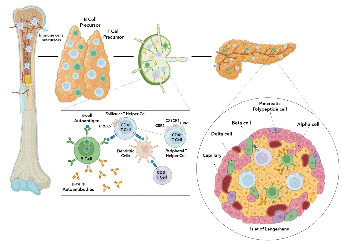
The pathogenesis of T1DM can be separated into a series of phases that starts with the identification of autoantibodies proceeding to cell death, dysglycemia, and hyperglycemiarelated symptoms [9]. The cause of cell-targeted autoimmunity involves a confluence of genetic and environmental variables that either initiate or facilitate the immune reaction against the cells [10]. The definitive cause of T1DM has not been determined; the variability of the condition is a challenging element when identifying disease-inducing variables. The numerous particular autoantibodies leading to autoimmunity are linked with diverse genetic susceptibility factors that reflect this variation [11,12]. T1DM is characterized by selective involvement of βcells without obvious pathogenic abnormalities of other Langerhans cells, such as α-cells, δ-cells, and pancreatic polypeptide cells [13]. High insulin autoantibody levels indicate early diabetes development in animals and young patients [14]. Therefore, mutations in the insulin gene promoter probably lead to a thymus with impaired central tolerance and the release of insulin-reactive T cells that direct the growth of autoreactive B cells [15]. Self-antigens secreted by damaged β cells are acquired by antigen-presenting cells (APCs), which then transport these materials to pancreatic lymphatic system and are passed on to autoreactive T lymphocytes. These T cells are uncontrolled components that are activated by genetically determined thymic deletion failures in combination with inadequacies in processes intended to potentiate peripheral immunological tolerance [16]. Following the inadequacies in central and peripheral tolerance, follicular T helper cells specific for islet antigens are generated in the pancreatic lymphatic system and assist autoreactive B cells in producing islet autoantibodies. Peripheral T helper cells generate CXCL13, which draws B cells towards inflamed islets, and interleukin (IL)-21, which enhances B-cell differentiation and leads to local autoantibody synthesis. Activated B cells generate cytokines and can operate as APCs to encourage increased immunological activation [17]. As a result of thymic plasma cell autoantibodies targeting certain medullary thymic epithelial cells for antibody-mediated apoptosis, the production of T cells is increased, bypassing negative selection and accelerating the course of T1DM [18].
B- and T-cell precursors are further developed in the thymus after formation in the bone marrow (Fig. 1) [19]. Beta cells and other islet cells produce type I interferon, such as interferon, which prompts the recruitment of immune cells. One of the initial cells to react are macrophages, which are also the major cell type that produces tumor necrosis factors (TNFs) [20]. Macrophages, acting as APCs, present autoantigens to the lymphatic system. Naive autoantigen-specific T lymphocytes in pancreatic lymph nodes identify islet molecular components from injured cells presented by APCs from pancreatic cells. CD4+, CD8+, and dendritic cells engage with activated B cells. T lymphocytes recognize autoantigens and eventually evade to the periphery [21,22]. Cell-specific T lymphocytes are activated by antigen presentation by dendritic cells and B cells. Autoreactive CD8+ and CD4+ T cells play significant roles in β-cell destruction by recognizing biomolecules of β-cell antigens such as precursor preproinsulin, tyrosine phosphatase-like insulinoma antigen, islet-specific glucose-6-phosphate catalytic subunit-related protein, glutamic acid decarboxylase-65, and zinc transport [23-25]. Additionally, B lymphocytes exposed to cell autoantigens produce islet-targeting autoantibodies. CXCR5+ antigen-specific follicular T helper cells promote autoreactive B cells to produce islet autoantibodies. Upon transition into peripheral T helper cells, a subpopulation of follicular T helper cells inhibits the expression CXCR5 and phosphorylates chemokine receptors CCR2, CCR5, and CX3CR1 [17,22]. Immunologic components, which invade the islet of Langerhans and damage insulin-producing cells, produce an inflammatory setting resembling insulitis. This prompts and expedites the establishment of T1DM by enhancing the susceptibility of the immune system to islet antigens carried by human leukocyte antigen class I molecules [26,27]. Immunological control imposed by regulatory T (Treg) cells and programmed cell death protein 1 ligation protects β-cells against autoimmune β-cell apoptosis. Insufficient immunological control can cause autoreactive T cells to initiate an autoimmune attack in T1DM, especially if these cells are activated by β-cells [26]. The destruction of insulinproducing β cells in the islet of Langerhans results in progressive hyperglycemia [28].

Current prevention strategies for T1DM

Several T1DM prevention strategies have been tested in clinical trials (Table 1) [29-41]. Although several studies have found the potential effects of delaying T1DM onset, short effective durations and detrimental toxicities pose challenges in long-term administration. Diabetic pharmacological drugs function by targeting the major signal transduction pathways associated with diabetes pathogenesis. The other pillars of diabetic treatment are nonpharmacological methods including nutritional adjustment, physical activity, and microbiotabased therapy [42]. Both hydrolyzed infant formula and glutenfree diet showed no significant effects in preventing the onset of T1DM [29,30]. According to several immunological studies, early childhood encounter with complex specific proteins may raise the incidence of β-cell autoimmunity in biologically vulnerable individuals [43,44]. Nevertheless, one study found that cow's milk protein plays no role in autoimmunity [29]. Delaying gluten introduction in children also plays no role in the onset of T1DM [30]. The use of vitamins such as vitamin B3, C, D, and E was to no avail [31-33].
The majority of randomized clinical studies examining type 1 diabetes-modifying medication have been undertaken in patients who had been clinically diagnosed with T1DM. Disease-modifying medications such as anti-CD3 monoclonal antibody, rituximab, abatacept, and alefacept substantially maintained insulin release while also exhibiting immunologic impacts [45-47]. Only a few preventive clinical studies are available due to the challenges in sample selection. Patients with T1DM undergo lifetime exogenous insulin replacement treatment as first-line therapy from the moment of diagnosis [48]. Insulin does not prevent or delay the onset of T1DM. Two clinical trials that differed in the assay method for autoantibody detection, micro insulin autoantibody assay versus radioimmunoassay, showed no benefit of oral insulin in preventing T1DM [34,35]. Moreover, the use of alum-formulated glutamate decarboxylase also had no significant effects [36].
TNF-α is a cytokine implicated in the inflammatory process that is generated by activated macrophages, CD4+ lymphocytes, and natural killer cells. TNF-α production may cause loss of pancreatic β cells, leading to the onset of T1DM [49]. Despite the lack of distinction between the groups in the concentration of glycated hemoglobin, the TNF-α inhibitor golimumab promoted improved indigenous insulin levels and decreased the need for insulin therapy when compared to placebo [37]. Moreover, the kinase inhibitor imatinib showed promising results in only the early stages of administration [38]. By addressing insulin resistance and endothelial dysfunction, tyrosine kinase inhibitors exhibit antihyperglycemic properties that have the potential to alleviate or prevent T1DM and type 2 diabetes mellitus [50]. In nonobese diabetic (NOD) mice, type 1 diabetes can be prevented but not reversed with a modest dosage of sorafenib (10 mg/kg/day). However, when sorafenib is administered at high doses (50 mg/kg/day), the drug causes preexisting type 1 diabetes in NOD mice to reemerge. This narrow therapeutic window may be attributed to the inhibition of T helper 1 (Th1) and Tc1 cells in the islets of Langerhans [51]. Similarly, the implementation of the immunosuppressant azathioprine immunosuppressant shows reconstructive insulin production but only in a transient manner [39]. T-celldirected immunosuppression only momentarily prevents the deterioration of cellular functions. The finding that T-celldirected immune intervention results in either no or only temporary retention of cellular functions shows that this therapy does not substantially affect the pathogenesis of T1DM [52]. Other operative approaches such as fecal microbiota transplantation and pancreas transplant alone have proven beneficial in preventing the occurrence of T1DM [40,41]. Nonetheless, there remain concerns, such as the emergence of an instant bloodmediated inflammatory reaction after operative measures, loss of islet volume and density owing to ischemia, islet cell apoptosis, and adverse immunosuppression drug side effects [53].

Dual actions of GABA to delay T1DM onset

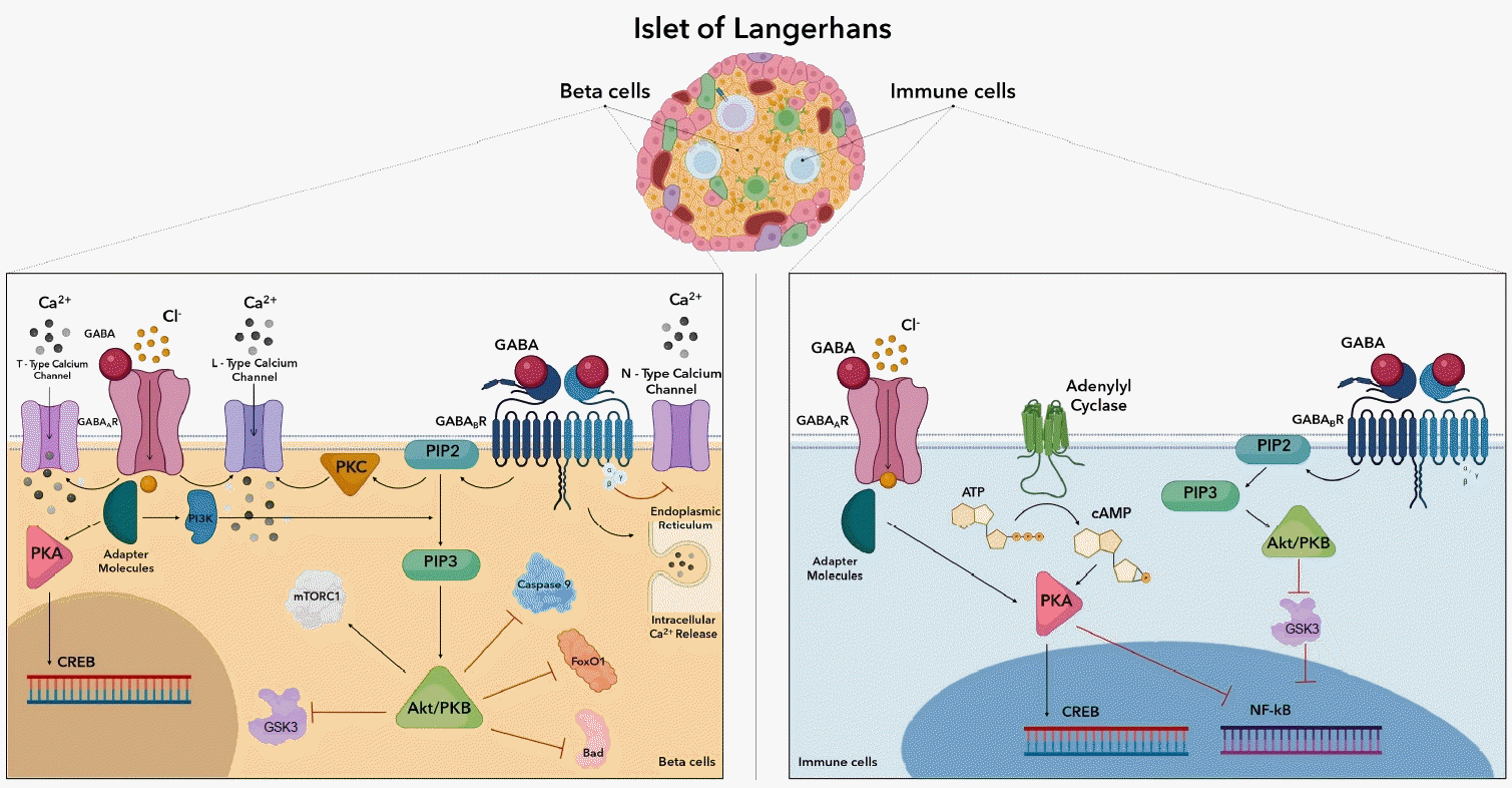
The GABA receptor is a multiunit postsynaptic receptor with 5 components arranged around a central pore. The GABA receptor has 2 classes, GABAA and GABAB, characterized by distinct toxicological, electrophysiological, and biochemical features [54]. GABA plays a significant role in both β cells and immune cells (Fig. 2). GABA receptor-mediated signaling involves adenylyl cyclase, voltage-gated Ca2+ channels, and G protein-activated inwardly rectifying K+ channels [55]. Activation of GABAA receptors leads to an increase in calcium ions through L- and T-type voltage-gated calcium channels. For β-cell GABAB receptors, activation opens potassium channels, releases Ca2+ from intracellular storage, activates protein kinase A (PKA), and stimulates cyclic adenosine monophosphate(cAMP)-response element binding protein (CREB) through a Ca2+-dependent pathway [56]. During maturation, GABAB receptor activation elevates calcium in specific neurons via protein kinase C stimulation [57].
GABAB receptor activation facilitates long-lasting (L-type) current and reduces current through N-type channels, promoting L-type calcium channels current via a non-Gi/o G protein. The Ca2+-dependent signaling pathway triggers phosphoinositide 3-kinase (PI3K)/phosphoinositidedependent kinase (Akt) signaling in response to calcium ion influx [58]. In human β cells, GABA-induced Ca2+ activates the Akt pathway [22]. PI3K catalyzes PIP3 (phosphatidylinositol (3,4,5)-trisphosphate) production, attracting signaling molecules with pleckstrin homology domains, such as protein kinase B and Akt [59]. Induced Akt regulates the phosphorylation of multiple downstream effectors, including glycogen synthase kinase 3 (GSK3), caspase-9, mammalian target of rapamycin complex 1 (mTORC1), Bcl-2-associated death promoter (Bad), and forkhead box protein O 1 (FoxO1) [60]. Akt directly controls cell viability by suppressing pro-apoptotic pathways such as the Bad and FoxO1 transcription factors. FoxO1 proteins either stimulate or repress the transcription of target genes. By modifying the transcription of pancreatic and duodenal homeobox-1 (PDX-1), the PI3K/AKT/FoxO1 signaling pathway can control insulin production [61]. Phosphorylation of Bad also inhibits the apoptotic effects formerly induced [62]. Caspase 9 is a crucial activator of the mitochondrial signaling pathways and a critical pro-apoptotic regulatory protein downstream of the PI3K/Akt pathway. The PI3K/Akt signaling pathway prevents apoptosis by suppressing caspase 9 signal transduction [63]. mTORC1 supports anabolic growth by stimulating protein, nucleotide, and lipid production while inhibiting catabolic mechanisms like autophagy through inhibition of Unc-51-like kinase 1 and transcription factor EB. By stimulation of its downstream targets ribosomal protein S6 kinase beta-1 and eukaryotic translation initiation factor 4E-binding protein 1, mTORC1 stimulates protein synthesis. The PI3K/mTORC1 pathway promotes β-cell development in human islets, and the addition of GABAA caused an increased β-cell area and multiplication [64].
GABAergic stimulation directly affects APCs, reduces MAPK signals, and reduces later adaptive inflammatory reactions to manage active autoimmune disorder [65]. Among T1DM patients, GABA controls the concentration-dependent production of 47 pro- and anti-inflammatory cytokines [66]. GABA may modify the immune system response to pathogens and play a role in autoimmune conditions such multiple sclerosis, type 1 diabetes, and rheumatoid arthritis. Several clinical trials have proven that GABA projects a variety of impacts on the immune system, including activating or suppressing cytokine release, altering cell proliferation, and influencing cell migration [67]. Specific depolarizing channel activation occurs in immune cells as most have ligand-specific gates, such as glutamate receptors and GABAA receptors, which have been found to affect both L- and T-type calcium channels [68]. Adenylate cyclase is a class of enzymes that catalyzes the synthesis of cAMP from adenosine triphosphate and is presumably controlled by a component of the Ca2+-signaling cascade [69]. Similarly, by means of intermediate processes involving intracellular second messengers, a GABAB receptor agonist upregulated basal resting adenylyl cyclase function and facilitated cAMP production. Additionally, cAMP is a significant down-regulator of T-cell function by reducing T-cell immunological activity via the cAMP/PKA/C-terminal Src kinase/lymphocyte-specific protein tyrosine kinase signaling cascade [70]. The transcription factor CREB is generated by PKA. Cell growth, maturation, and survival are all regulated by CREB. CREB regulates T-cell activity and promotes Treg production and stability. Moreover, CREB has been demonstrated to promote transcription of CRE-containing immune-related cytokines such as IL-2, IL-6, IL-10, and TNF-α [70]. Additionally, PKA-dependent activation has the potential to downregulate nuclear factor kappa-lightchain-enhancer of activated B cells (NF-κB) activity. Several studies have proven that increased NF-κB activity is associated with the pathogenesis of autoimmunity, and downregulating this transcription factor would pose therapeutic benefits [71]. The stimulation of GABAB receptor resulted in the suppression of GSK3 expression, which restricted NF-κB activity [72]. Given its numerous roles in cell physiology in T1DM, cAMP has a wide range of modulatory associations on a spectrum of immune cells (Table 2) [66,73-79]. Several studies show that in children who eventually developed T1DM, dysregulation of the GABA metabolism and other circulating metabolites occurred before pancreatic islets autoimmunity [80]. Autoimmunity may therefore be a late reaction to the preceding metabolic abnormalities; thus, adhering to metabolic needs could be an effective delaying or preventive measure.

GABAB receptor agonist in children

Previous studies suggested that by stimulating cell proliferation and survival through the activation of the PI3-K/Akt pathway, GABA is able to delay and reverse the onset of T1D [81,82]. GABA is also able to exert immunoinhibitory effects, precisely by suppressing the activity of Th1 cells against islets, cytotoxic cells; suppressing lymphocyte proliferation and the activation of NF-κB; and increasing the number of Treg cells [83]. GABA is also able to inhibit the secretion of glucagon and has been suggested to have the ability to induce transdifferentiation of α-like cells into β-like cells by acting on the GABAA receptor with the assistance of gephyrin. This results in islet hyperplasia [84]. An experiment showed that GABA-induced differentiation of pancreatic stem cells into insulin-producing islet cells; this was proven by the positive immunostaining results of PDX-1 [85]. PDX-1 and NKx6.1 are 2 of the major markers in β cell lineage investigations [86].
Multiple studies conducted in vivo have shown a potentially positive effect of GABA administration in inducing remission or delaying the onset of T1DM (Table 3). A study in female NOD mice has proven the effect to be dose-dependent as lower doses of lesogaberan (0.025 mg/mL in drinking water) did not produce any significant improvement. The therapeutic effect between doses of 0.25 and 0.75 mg/mL showed little variance [87]. Other studies also identified a dose-dependent relationship between GABA administration and delayed onset of T1DM [88,89]. NOD/SCID mice were given GABA treatment prior to the transfusion of diabetogenic splenic cells. These results show that GABA prevents adoptive transfer of T1DM. GABA also delays T1DM progression in 6-week-old prediabetic NOD mice with autoimmunity. After treatment, at 40 weeks of age, none of the mice were found to be hyperglycemic. The research also suggested that GABA suppresses Th1 activity against islet antigens and consequently inhibits the proliferation of autoreactive T cells [88]. Another study conducted in NSG mice induced with diabetes using streptozotocin showed that oral GABA administration stimulates increase in the mass of β cells, proven by an approximate fivefold increase of β-cell multiplication. The study also concluded that GABA treatment increases insulin levels and exerts protective effects against apoptosis of β cells [56]. Similar conclusions were drawn from another study conducted in NOD/SCID mice in which GABA enhanced islet β-cell proliferation and inhibited β-cell apoptosis [8]. A different study conducted in 8-week-old male FVB mice showed that oral administration of muscimol(GABAA receptor agonist) and baclofen (GABAB receptor agonist) increased proliferation of β cells by 27%±10% and 47%±16% respectively. Baclofen also increased proliferation of α-cells by 72%±21% [90]. Increased expression of PDX-1 and NKx6.1 were found in another study, indicating increased proliferation of β cells. Beta-cell rate of apoptosis also decreased to around 0.5% as compared to 1.5% in untreated mice and GABA and insulin levels increased [86]. A clinical trial proposed to examine the safety and efficacy of GABA in delaying the onset or progression of T1DM in children is currently undergoing phase I clinical trial [91].

Adverse effects and limitations of GABA treatment

The highest concentration of GABA is found in approximately 30% of the brain's neurons and is found in the pancreas at similar levels [92]. GABA is a cerebral neurotransmitter inhibitor which makes CNS depression a frequent adverse effect (AE) [93]. However, oral administration of GABA has been proven to be safe and effective in vivo, oral administration leads to peripheral action and prevents crossing of the blood-brain barrier [83].
The United States Pharmacopeia conducted a review on the safety of GABA. GABA treatment is associated with a brief mild drop (<10%) in blood pressure, abdominal discomfort, headaches, drowsiness, and a brief burning sensation in the throat. All AEs reported ranged from mild to moderate. Multiple clinical studies on GABA use have reported no serious AEs. Importantly, GABA treatment is discouraged in pregnant and breastfeeding women as GABA may cause an increase in growth hormone and prolactin levels. However, no specific studies have mentioned negative effects [94]. That sudden cessation of therapy may lead to withdrawal symptoms is also important to note [95].

Conclusions

Despite substantial advancements in technology, knowledge gaps remain in the understanding and management of T1DM. Multiple preclinical and clinical studies have been conducted to test the potential use of GABA in delaying T1DM, especially in high-risk groups such as children with genetic predisposition for T1DM. GABA exerts dual action on both islet β cells and immune cells. The studies discussed in this article have shown regenerative and protective effects on islet β cells. GABA induces proliferation of β cells and inhibits β-cell apoptosis. No serious AEs related to GABA treatment were reported. These GABA study results create optimism in its potential to delay the onset of T1DM in children.

Notes

Conflicts of interest

No potential conflict of interest relevant to this article was reported.

Funding

This study received no specific grant from any funding agency in the public, commercial, or not-for-profit sectors.

Author contribution

Conceptualization: JCS; Formal analysis: MRS; Methodology: BGL; Visualization: BGL; Writing - original draft: JCS, BGL, MRS; Writing - review & editing: JCS, BGL, MRS

ACKNOWLEDGMENTS

The authors would like to express gratitude towards every party involved in the generation of this manuscript.

Fig. 1.
Type 1 diabetes mellitus autoimmunity pathophysiology.
apem-2346184-092f1.tif
Fig. 2.
Dual pharmacodynamics of GABA in β-cells and immune cells. GABA, gamma-aminobutyric acid; GABAA R, GABA subtype A receptor; GABAB R, GABA subtype B receptor; PI3K, phosphoinositide 3-kinase; PKC, protein kinase C; PIP2, phosphatidylinositol (4,5)-bisphosphate; PIP3, phosphatidylinositol (3,4,5)-trisphosphate; mTORC1, mammalian target of rapamycin complex 1; PKB, protein kinase B; PKA, protein kinase A; cAMP, cyclic adenosine monophosphate; CREB, cAMP-response element binding; GSK3, glycogen synthase kinase 3; ATP, adenosine triphosphate; NF-κB, nuclear factor kappa-light-chain-enhancer of activated B cells.
apem-2346184-092f2.tif
Table 1.
Clinical trials in delaying the onset of T1DM
Target Agent Findings Reference
Diet Hydrolyzed infant formula No reduction in T1DM incidence rate after 11.5 years [29]
Gluten-free diet Did not significantly lower the likelihood of islet autoimmunity in children who are genetically susceptible. [30]
Vitamins Niacinamide (vitamin B3) Treatment with niacinamide did not significantly reduce or postpone the onset of T1DM. [31]
Cholecalciferol (vitamin D) Neither vitamin D consumption nor 25(OH)D levels were linked to the likelihood of IA or the development of type 1 diabetes. [32]
Ascorbic acid and tocopherol (vitamins C and E) Supplementation of vitamins C and E antioxidants did not enhance endothelial function, endothelial colony forming cells, and other unconventional risk factors. [33]
Hormones Oral insulin Over 2.7 years, oral insulin at a dosage of 7.5 mg/day, compared to placebo, did not prevent or postpone the onset of T1DM. [34,35]
Enzyme Alum-formulated glutamate decarboxylase No influence on the development of type 1 diabetes [36]
TNF-α Inhibitor Golimumab The mean glycated hemoglobin levels were similar among the groups, but results indicated that golimumab was linked to an increase and longer duration of partial remission. [37]
Kinase inhibitors Imatinib Slowed decline of β-cells up to 12 months. This benefit did not last for 24 months, and 71% of patients experienced grade 2 adverse side effects. [38]
Immune suppressant Azathioprine Azathioprine alone has little effect on the remission stage despite initial impacts on endogenous insulin production. [39]
Operative Fecal microbiota transplantation Plasma 1-arachidonoyl-GPC, Desulfovibrio piger inhibits autoimmunity in T1DM, impacting CXCR3+ T cells to an extent [40]
Pancreas transplant alone (PTA) PTA is supported as a viable treatment option by long-term effects on survival rates, transplant functionality, and the native kidneys. [41]

T1DM, type 1 diabetes mellitus; 25(OH)D, 25-hydroxyvitamin D; TNF, tumor necrosis factor.

Table 2.
Immunoregulatory effects of GABA
Immune cells GABA outcome Reference
Macrophage Downregulate proinflammatory IL-12 and IL-6 production [73]
T cells Decrease proliferation of CD4+ and downregulate IFN-γ, Flt3L, TRAIL, TNF-β, PD-L1, and IL-10 production [66,74]
Natural killer cells Decreased cytotoxicity and cytokine release in vitro and target cell lysis inhibition by perforin and CD95 ligand [75-77]
Dendritic cells Mitogen-activated protein kinases are increased, cytokine actions are modulated, and production of anti-inflammatory cytokine IL-10 is encouraged [78,79]
B Cells The immunomodulatory effects of GABAergic constituents in B cells and granulocytes are poorly understood. [75]

GABA, gamma-aminobutyric acid; IL, interleukin; IFN, interferon; Flt3L, Fms-like tyrosine kinase 3 ligand; TRAIL, TNF-related apoptosis inducing ligand; TNF, tumor necrosis factor; PD-L1, programmed death ligand 1.

Table 3.
Effects of GABA in vivo
Subject Route Dosage Duration Results Reference
Female NOD mice Oral 0.025, 0.08, 0.25, 0.75 mg/mL of lesogaberan (AZD3355) Up to 28 weeks post onset of T1DM 0.025 mg/mL: no significant improvement [87]
0.08 mg/mL: around 50% experienced transient remission
0.25 or 0.75 mg/mL: remission for an average duration of 4.4 and 5.8 weeks consecutively
6 and 20 w eeks old female NOD or SCID mice Pellet implantation 600 μg/day 90 Days Delayed onset of T1DM, lo w ered incidence of T1DM, no signs of hyperglycemia at 40 weeks of age [88]
Male NOD-SCID-γ (NSG) mice Oral 6 mg/mL 5 Weeks GABA administration enhanced β-cell proliferation by a factor of about five [56]
2.5 to 10 months old wild-type mice Intraperitoneal (IP) injection 250 μg/kg/day 1–6 Months Doubled number of islets in mice, direc tly propor tional increase of insulin-producing cells with duration of treatment, increase in β-like cell mass [89]
NOD/SCID mice Oral 6 mg/mL 12 Days Increases proliferation and restricts apoptosis of β cells [8]
Six adult male subjects with chronic T1DM Oral 200, 600, 1,200 mg/day 11 Days No severe adverse effects (AEs) were reported, while mild and transient AEs occurred [5]
Eight-week-old male FVB mice Oral 6 mg/mL of muscimol or baclofen 6 Weeks Muscimol: increased β-cell multiplication by 27%±10% [90]
Baclofen: increased β-cell multiplication by 47%±16% and α-cell multiplication by 72%±21%
Male NSG mice Oral 6 mg/mL 10 Weeks Increased expression of pancreatic and duodenal homeobox-1 and NK6 homeobox 1, reduced apoptosis of β-cells to around 0.5%, increased number of β-cells and insulin levels and enhanced glucose homeostasis [86]

GABA, gamma-aminobutyric acid; NOD, nonobese diabetic; T1DM, type 1 diabetes mellitus; SCID, severely combined immunodeficient.

References

1. Kahanovitz L, Sluss PM, Russell SJ. Type 1 diabetes - a clinical perspective. Point Care 2017;16:37–40.
crossref pmid pmc
2. Gregory GA, Robinson TIG, Linklater SE, Wang F, Colagiuri S, de Beaufort C, et al. Global incidence, prevalence, and mortality of type 1 diabetes in 2021 with projection to 2040: a modelling study. Lancet Diabetes Endocrinol 2022;10:741–60.
crossref pmid
3. Rafferty J, Stephens JW, Atkinson MD, Luzio SD, Akbari A, Gregory JW, et al. A retrospective epidemiological study of type 1 diabetes mellitus in Wales, UK between 2008 and 2018. Int J Popul Data Sci 2021;6:1387.
crossref pmid pmc
4. Karras SN, Koufakis T, Zebekakis P, Kotsa K. Pharmacologic adjunctive to insulin therapies in type 1 diabetes: the journey has just begun. World J Diabetes 2019;10:234–40.
crossref pmid pmc
5. Espes D, Liljebäck H, Hill H, Elksnis A, Caballero-Corbalan J, Birnir B, et al. GABA induces a hormonal counterregulatory response in subjects with long-standing type 1 diabetes. BMJ Open Diabetes Res Care 2021;9:e002442.
crossref pmid pmc
6. Rachdi L, Maugein A, Pechberty S, Armanet M, Hamroune J, Ravassard P, et al. Regulated expression and function of the GABAB receptor in human pancreatic beta cell line and islets. Sci Rep 2020;10:13469.
crossref pmid pmc
7. Badri H, Gibbard C, Denton D, Satia I, Al-Sheklly B, Dockry RJ, et al. A double-blind randomised placebocontrolled trial investigating the effects of lesogaberan on the objective cough frequency and capsaicin-evoked coughs in patients with refractory chronic cough. ERJ Open Res 2022;8:00546–2021.
crossref pmid pmc
8. Tian J, Dang H, Hu A, Xu W, Kaufman DL. Repurposing Lesogaberan to promote human islet cell survival and β-cell replication. J Diabetes Res 2017;2017:6403539.
crossref pmid pmc
9. Insel RA, Dunne JL, Atkinson MA, Chiang JL, Dabelea D, Gottlieb PA, et al. Staging presymptomatic type 1 diabetes: a scientific statement of JDRF, the Endocrine Society, and the American Diabetes Association. Diabetes Care 2015;38:1964–74.
crossref pmid pmc
10. Katsarou A, Gudbjörnsdottir S, Rawshani A, Dabelea D, Bonifacio E, Anderson BJ, et al. Type 1 diabetes mellitus. Nat Rev Dis Prim 2017;3:17016.
crossref pmid
11. Yahaya T, Adedayo T. Genes predisposing to type 1 diabetes mellitus and pathophysiology: a narrative review. Med J Indones 2020;29:100–9.
crossref
12. Krischer JP, Lynch KF, Lernmark Å, Hagopian WA, Rewers MJ, She JX, et al. Genetic and environmental interactions modify the risk of diabetes-related autoimmunity by 6 years of age: the TEDDY study. Diabetes Care 2017;40:1194–202.
crossref pmid pmc
13. Zaccardi F, Webb DR, Yates T, Davies MJ. Pathophysiology of type 1 and type 2 diabetes mellitus: a 90-year perspective. Postgrad Med J 2016;92:63–9.
crossref pmid
14. Steck AK, Johnson K, Barriga KJ, Miao D, Yu L, Hutton JC, et al. Age of islet autoantibody appearance and mean levels of insulin, but not GAD or IA-2 autoantibodies, predict age of diagnosis of type 1 diabetes: diabetes autoimmunity study in the young. Diabetes Care 2011;34:1397–9.
pmid pmc
15. Fousteri G, Ippolito E, Ahmed R, Hamad ARA. Beta-cell specific autoantibodies: are they just an indicator of type 1 diabetes? Curr Diabetes Rev 2017;13:322–9.
crossref pmid pmc
16. Bluestone JA, Herold K, Eisenbarth G. Genetics, pathogenesis and clinical interventions in type 1 diabetes. Nature 2010;464:1293–300.
crossref pmid pmc
17. Vandamme C, Kinnunen T. B cell helper T cells and type 1 diabetes. Scand J Immunol 2020;92:e12943.
crossref pmid pmc
18. Greaves RB, Chen D, Green EA. Thymic B cells as a new player in the type 1 diabetes response. Front Immunol 2021;12:772017.
crossref pmid pmc
19. Perera J, Huang H. The development and function of thymic B cells. Cell Mol Life Sci 2015;72:2657–63.
crossref pmid pmc
20. Colli ML, Szymczak F, Eizirik DL. Molecular footprints of the immune assault on pancreatic beta cells in type 1 diabetes. Front Endocrinol (Lausanne) 2020:11:568446.

21. Zajec A, Trebušak Podkrajšek K, Tesovnik T, Šket R, Čugalj Kern B, Jenko Bizjan B, et al. Pathogenesis of type 1 diabetes: established facts and new insights. Genes (Basel) 2022;13:706.
crossref pmid pmc
22. Ilonen J, Lempainen J, Veijola R. The heterogeneous pathogenesis of type 1 diabetes mellitus. Nat Rev Endocrinol 2019;15:635–50.
crossref pmid
23. Culina S, Lalanne AI, Afonso G, Cerosaletti K, Pinto S, Sebastiani G, et al. Islet-reactive CD8+ T cell frequencies in the pancreas, but not in blood, distinguish type 1 diabetic patients from healthy donors. Sci Immunol 2018;3:eaao4013.
crossref pmid
24. Yu W, Jiang N, Ebert PJ, Kidd BA, Müller S, Lund PJ, et al. Clonal deletion prunes but does not eliminate self-specific αβ CD8(+) T lymphocytes. Immunity 2015;42:929–41.
crossref pmid pmc
25. Gonzalez-Duque S, Azoury ME, Colli ML, Afonso G, Turatsinze JV, Nigi L, et al. Conventional and neo-antigenic peptides presented by β cells are targeted by circulating naïve CD8+ T cells in type 1 diabetic and healthy donors. Cell Metab 2018;28:946–60.e6.
crossref pmid
26. Roep BO, Thomaidou S, van Tienhoven R, Zaldumbide A. Type 1 diabetes mellitus as a disease of the β-cell (do not blame the immune system?). Nat Rev Endocrinol 2021;17:150–61.
crossref pmid
27. Coppieters KT, Dotta F, Amirian N, Campbell PD, Kay TW, Atkinson MA, et al. Demonstration of islet-autoreactive CD8 T cells in insulitic lesions from recent onset and longterm type 1 diabetes patients. J Exp Med 2012;209:51–60.
crossref pmid pmc
28. Willcox A, Gillespie KM. Histology of type 1 diabetes pancreas. Methods Mol Biol 2016;1433:105–17.
crossref pmid
29. Writing Group for the TRIGR Study Group. Effect of hydrolyzed infant formula vs conventional formula on risk of type 1 diabetes: the TRIGR randomized clinical trial. JAMA 2018;319:38–48.
pmid pmc
30. Hummel S, Pflüger M, Hummel M, Bonifacio E, Ziegler AG. Primary dietary intervention study to reduce the risk of islet autoimmunity in children at increased risk for type 1 diabetes: the BABYDIET study. Diabetes Care 2011;34:1301–5.
pmid pmc
31. Lampeter EF, Klinghammer A, Scherbaum WA, Heinze E, Haastert B, Giani G, et al. The Deutsche Nicotinamide Intervention Study: an attempt to prevent type 1 diabetes. DENIS Group. Diabetes 1998;47:980–4.
crossref pmid
32. Simpson M, Brady H, Yin X, Seifert J, Barriga K, Hoffman M, et al. No association of vitamin D intake or 25-hydroxyvitamin D levels in childhood with risk of islet autoimmunity and type 1 diabetes: the Diabetes Autoimmunity Study in the Young (DAISY). Diabetologia 2011;54:2779–88.
crossref pmid pmc
33. Cazeau RM, Huang H, Bauer JA, Hoffman RP. Effect of vitamins C and E on endothelial function in type 1 diabetes mellitus. J Diabetes Res 2016;2016:3271293.
crossref pmid
34. Skyler JS, Krischer JP, Wolfsdorf J, Cowie C, Palmer JP, Greenbaum C, et al. Effects of oral insulin in relatives of patients with type 1 diabetes: the diabetes prevention trial--type 1. Diabetes Care 2005;28:1068–76.
pmid
35. Krischer JP, Schatz DA, Bundy B, Skyler JS, Greenbaum CJ. Effect of oral insulin on prevention of diabetes in relatives of patients with type 1 diabetes: a randomized clinical trial. JAMA 2017;318:1891–902.
crossref pmid pmc
36. Elding Larsson H, Lundgren M, Jonsdottir B, Cuthbertson D, Krischer J. Safety and efficacy of autoantigen-specific therapy with 2 doses of alum-formulated glutamate decarboxylase in children with multiple islet autoantibodies and risk for type 1 diabetes: a randomized clinical trial. Pediatr Diabetes 2018;19:410–9.
crossref pmid
37. Quattrin T, Haller MJ, Steck AK, Felner EI, Li Y, Xia Y, et al. Golimumab and beta-cell function in youth with newonset type 1 diabetes. N Engl J Med 2020;383:2007–17.
crossref pmid
38. Gitelman SE, Bundy BN, Ferrannini E, Lim N, Blanchfield JL, DiMeglio LA, et al. Imatinib therapy for patients with recent-onset type 1 diabetes: a multicentre, randomised, double-blind, placebo-controlled, phase 2 trial. lancet Diabetes Endocrinol 2021;9:502–14.
pmid pmc
39. Cook JJ, Hudson I, Harrison LC, Dean B, Colman PG, Werther G, et al. Double-blind controlled trial of azathioprine in children with newly diagnosed type i diabetes. Diabetes 1989;38:779–83.
crossref pmid
40. de Groot P, Nikolic T, Pellegrini S, Sordi V, Imangaliyev S, Rampanelli E, et al. Faecal microbiota transplantation halts progression of human new-onset type 1 diabetes in a randomised controlled trial. Gut 2021;70:92–105.
crossref pmid
41. Boggi U, Baronti W, Amorese G, Pilotti S, Occhipinti M, Perrone V, et al. Treating type 1 diabetes by pancreas transplant alone: a cohort study on actual long-term (10 years) efficacy and safety. Transplantation 2022;106:147–57.
crossref pmid
42. Wang X, Kang J, Liu Q, Tong T, Quan H. Fighting diabetes mellitus: pharmacological and non-pharmacological approaches. Curr Pharm Des 2020;26:4992–5001.
crossref pmid
43. Ziegler AG, Schmid S, Huber D, Hummel M, Bonifacio E. Early infant feeding and risk of developing type 1 diabetesassociated autoantibodies. JAMA 2003;290:1721–8.
crossref pmid
44. Knip M, Virtanen SM, Seppä K, Ilonen J, Savilahti E, Vaarala O, et al. Dietary intervention in infancy and later signs of beta-cell autoimmunity. N Engl J Med 2010;363:1900–8.
crossref pmid pmc
45. Orban T, Bundy B, Becker DJ, Dimeglio LA, Gitelman SE, Goland R, et al. Costimulation modulation with abatacept in patients with recent-onset type 1 diabetes: followup 1 year after cessation of treatment. Diabetes Care 2014;37:1069–75.
crossref pmid pmc
46. Rigby MR, Harris KM, Pinckney A, DiMeglio LA, Rendell MS, Felner EI, et al. Alefacept provides sustained clinical and immunological effects in new-onset type 1 diabetes patients. J Clin Invest 2015;125:3285–96.
crossref pmid pmc
47. Pescovitz MD, Greenbaum CJ, Bundy B, Becker DJ, Gitelman SE, Goland R, et al. B-lymphocyte depletion with rituximab and β-cell function: two-year results. Diabetes Care 2014;37:453–9.
crossref pmid pmc
48. Janež A, Guja C, Mitrakou A, Lalic N, Tankova T, Czupryniak L, et al. Insulin therapy in adults with type 1 diabetes mellitus: a narrative review. Diabetes Ther Res Treat Educ diabetes Relat Disord 2020;11:387–409.
crossref
49. Qiao YC, Chen YL, Pan YH, Tian F, Xu Y, Zhang XX, et al. The change of serum tumor necrosis factor alpha in patients with type 1 diabetes mellitus: a systematic review and meta-analysis. PLoS One 2017;12:e0176157.
crossref pmid pmc
50. Fountas A, Diamantopoulos LN, Tsatsoulis A. Tyrosine kinase inhibitors and diabetes: a novel treatment paradigm? Trends Endocrinol Metab 2015;26:643–56.
crossref pmid
51. Zeng Q, Song J, Wang D, Sun X, Xiao Y, Zhang H, et al. Identification of sorafenib as a treatment for type 1 diabetes. Front Immunol 2022;13:740805.
crossref pmid pmc
52. Kolb H, von Herrath M. Immunotherapy for type 1 diabetes: why do current protocols not halt the underlying disease process? Cell Metab 2017;25:233–41.
crossref pmid
53. Bottino R, Knoll MF, Knoll CA, Bertera S, Trucco MM. The future of islet transplantation is now. Front Med 2018;5:202.
crossref
54. Uusi-Oukari M, Korpi ER. Regulation of GABA(A) receptor subunit expression by pharmacological agents. Pharmacol Rev 2010;62:97–135.
crossref pmid
55. Dwyer TM. Chapter 4 - Chemical signaling in the nervous system. Haines DE, Mihailoff GAet al., editors. Fundamental neuroscience for basic and clinical applications (fifth edition). Amsterdam (Netherlands): Elsevier. 2018;pp 54–71.e1.

56. Purwana I, Zheng J, Li X, Deurloo M, Son DO, Zhang Z, et al. GABA promotes human β-cell proliferation and modulates glucose homeostasis. Diabetes 2014;63:4197–205.
crossref pmid
57. Karls A, Mynlieff M. GABAB receptors couple to Gαq to mediate increases in voltage-dependent calcium current during development. J Neurochem 2015;135:88–100.
crossref pmid pmc
58. Leng S, Xie F, Liu J, Shen J, Quan G, Wen T. LLGL2 increases Ca(2+) influx and exerts oncogenic activities via PI3K/AKT signaling pathway in hepatocellular carcinoma. Front Oncol 2021;11:683629.
crossref pmid pmc
59. Marat AL, Haucke V. Phosphatidylinositol 3-phosphates-at the interface between cell signalling and membrane traffic. EMBO J 2016;35:561–79.
crossref pmid pmc
60. Camaya I, Donnelly S, O'Brien B. Targeting the PI3K/Akt signaling pathway in pancreatic β-cells to enhance their survival and function: an emerging therapeutic strategy for type 1 diabetes. J Diabetes 2022;14:247–60.
pmid pmc
61. Zhang B, Sun P, Shen C, Liu X, Sun J, Li D, et al. Role and mechanism of PI3K/AKT/FoxO1/PDX-1 signaling pathway in functional changes of pancreatic islets in rats after severe burns. Life Sci 2020;258:118145.
crossref pmid
62. Dasari S, Tchounwou PB. Cisplatin in cancer therapy: molecular mechanisms of action. Eur J Pharmacol 2014;740:364–78.
crossref pmid pmc
63. Wang J, Liu H, Zhang X, Li X, Geng L, Zhang H, et al. Sulfated hetero-polysaccharides protect SH-SY5Y cells from H2O2-induced apoptosis by affecting the PI3K/Akt signaling pathway. Mar Drugs 2017;15:110.
crossref pmid pmc
64. Ardestani A, Lupse B, Kido Y, Leibowitz G, Maedler K. mTORC1 signaling: a double-edged sword in diabetic β cells. Cell Metab 2018;27:314–31.
crossref pmid
65. Bhat R, Axtell R, Mitra A, Miranda M, Lock C, Tsien RW, et al. Inhibitory role for GABA in autoimmune inflammation. Proc Natl Acad Sci 2010;107:2580–5.
crossref pmid pmc
66. Bhandage AK, Jin Z, Korol SV, Shen Q, Pei Y, Deng Q, et al. GABA regulates release of inflammatory cytokines from peripheral blood mononuclear cells and CD4+ T cells and is immunosuppressive in type 1 diabetes. EBioMedicine 2018;30:283–94.
crossref pmid pmc
67. Dionisio L, José De Rosa M, Bouzat C, Esandi Mdel C. An intrinsic GABAergic system in human lymphocytes. Neuropharmacology 2011;60:513–9.
crossref pmid
68. Bhandage AK, Jin Z, Hellgren C, Korol SV, Nowak K, Williamsson L, et al. AMPA, NMDA and kainate glutamate receptor subunits are expressed in human peripheral blood mononuclear cells (PBMCs) where the expression of GluK4 is altered by pregnancy and GluN2D by depression in pregnant women. J Neuroimmunol 2017;305:51–8.
crossref pmid
69. Halls ML, Cooper DM. Regulation by Ca2+-signaling pathways of adenylyl cyclases. Cold Spring Harb Perspect Biol 2011;3:a004143.
crossref pmid pmc
70. Wehbi VL, Taskén K. Molecular mechanisms for cAMPmediated immunoregulation in T cells - role of anchored protein kinase A signaling units. Front Immunol 2016;7:222.
crossref pmid pmc
71. Barnabei L, Laplantine E, Mbongo W, Rieux-Laucat F, Weil R. NF-κB: at the borders of autoimmunity and inflammation. Front Immunol 2021;12:716469.
crossref pmid pmc
72. Shu Q, Liu J, Liu X, Zhao S, Li H, Tan Y, et al. GABAB R/GSK-3β/NF-κB signaling pathway regulates the proliferation of colorectal cancer cells. Cancer Med 2016;5:1259–67.
pmid pmc
73. Reyes-García MG, Hernández-Hernández F, Hernández-Téllez B, García-Tamayo F. GABA (A) receptor subunits RNA expression in mice peritoneal macrophages modulate their IL-6/IL-12 production. J Neuroimmunol 2007;188:64–8.
crossref pmid
74. Wei M, Li L, Meng R, Fan Y, Liu Y, Tao L, et al. Suppressive effect of diazepam on IFN-gamma production by human T cells. Int Immunopharmacol 2010;10:267–71.
pmid
75. Bhandage AK, Barragan A. GABAergic signaling by cells of the immune system: more the rule than the exception. Cell Mol Life Sci 2021;78:5667–79.
crossref pmid pmc
76. Goto M, Murakawa M, Kadoshima-Yamaoka K, Tanaka Y, Inoue H, Murafuji H, et al. Phosphodiesterase 7A inhibitor ASB16165 suppresses proliferation and cytokine production of NKT cells. Cell Immunol 2009;258:147–51.
crossref pmid
77. Zhao W, Huang Y, Liu Z, Cao BB, Peng YP, Qiu YH. Dopamine Receptors modulate cytotoxicity of natural killer cells via cAMP-PKA-CREB signaling pathway. PLoS One 2013;8:e65860.
crossref pmid pmc
78. I Ciftci H, G Sierra R, Yoon CH, Su Z, Tateishi H, Koga R, et al. Serial femtosecond x-ray diffraction of HIV-1 gag MAIP6 microcrystals at ambient temperature. Int J Mol Sci 2019;20:1675.
crossref pmid pmc
79. Oger S, Méhats C, Dallot E, Cabrol D, Leroy MJ. Evidence for a role of phosphodiesterase 4 in lipopolysaccharidestimulated prostaglandin E2 production and matrix metalloproteinase-9 activity in human amniochorionic membranes. J Immunol 2005;174:8082–9.
crossref pmid
80. Lamichhane S, Sen P, Dickens AM, Alves MA, Härkönen T, Honkanen J, et al. Dysregulation of secondary bile acid metabolism precedes islet autoimmunity and type 1 diabetes. Cell Reports Med 2022;3:100762.
crossref
81. Yi Z, Waseem Ghani M, Ghani H, Jiang W, Waseem Birmani M, Ye L, et al. Gimmicks of gamma-aminobutyric acid (GABA) in pancreatic β-cell regeneration through transdifferentiation of pancreatic α- to β-cells. Cell Biol Int 2020;44:926–36.
crossref pmid
82. Soltani N, Qiu H, Aleksic M, Glinka Y, Zhao F, Liu R, et al. GABA exerts protective and regenerative effects on islet beta cells and reverses diabetes. Proc Natl Acad Sci 2011;108:11692–7.
crossref pmid pmc
83. Wang Q, Prud'homme G, Wan Y. GABAergic system in the endocrine pancreas: a new target for diabetes treatment. Diabetes Metab Syndr Obes Targets Ther 2015;8:79–87.
crossref
84. Weir GC, Bonner-Weir S. GABA signaling stimulates β cell regeneration in diabetic mice. Cell 2017;168:7–9.
crossref pmid
85. Ghani MW, Yi Z, Jiang W, Bin L, Cun LG, Birmany MW, et al. Gamma-aminobutyric acid (GABA) induced in vitro differentiation of rat pancreatic ductal stem cells into insulin-secreting islet-like cell clusters. Folia Biol (Praha) 2019;65:246–55.
pmid
86. Liu W, Lau HK, Son DO, Jin T, Yang Y, Zhang Z, et al. Combined use of GABA and sitagliptin promotes human β-cell proliferation and reduces apoptosis. J Endocrinol 2021;248:133–43.
crossref pmid
87. Tian J, Middleton B, Lee VS, Park HW, Zhang Z, Kim B, et al. GABA(B)-receptor agonist-based immunotherapy for type 1 diabetes in NOD mice. Biomedicines 2021;9:43.
crossref pmid pmc
88. Tian J, Lu Y, Zhang H, Chau CH, Dang HN, Kaufman DL. Gamma-aminobutyric acid inhibits T cell autoimmunity and the development of inflammatory responses in a mouse type 1 diabetes model. J Immunol 2004;173:5298–304.
pmid
89. Ben-Othman N, Vieira A, Courtney M, Record F, Gjernes E, Avolio F, et al. Long-term GABA administration induces alpha cell-mediated beta-like cell neogenesis. Cell 2017;168:73–85.e11.
crossref pmid
90. Untereiner A, Xu J, Bhattacharjee A, Cabrera O, Hu C, Dai FF, et al. Gamma-aminobutyric acid stimulates β-cell proliferation through the mTORC1/p70S6K pathway, an effect amplified by Ly49, a novel γ-aminobutyric acid type A receptor positive allosteric modulator. Diabetes Obes Metab 2020;22:2021–31.
pmid
91. Choat HM, Martin A, Mick GJ, Heath KE, Tse HM, McGwin GJ, et al. Effect of gamma aminobutyric acid (GABA) or GABA with glutamic acid decarboxylase (GAD) on the progression of type 1 diabetes mellitus in children: Trial design and methodology. Contemp Clin Trials 2019;82:93–100.
crossref pmid
92. Hagan DW, Ferreira SM, Santos GJ, Phelps EA. The role of GABA in islet function. Front Endocrinol (Lausanne) 2022;13:972115.
crossref pmid pmc
93. Allen MJ, Sabir S, Sharma S. GABA receptor. 2023 Feb 13. In: StatPearls [Internet]. Treasure Island (FL): StatPearls Publishing; Jan–.

94. Oketch-Rabah HA, Madden EF, Roe AL, Betz JM. United States Pharmacopeia (USP) safety review of gammaaminobutyric acid (GABA). Nutrients 2021;13:2742.
crossref pmid pmc
95. Janković SM, Dješević M, Janković SV. Experimental GABA A receptor agonists and allosteric modulators for the treatment of focal epilepsy. J Exp Pharmacol 2021;13:235–44.
pmid pmc