Ann Pediatr Endocrinol Metab Search

CLOSE


Ann Pediatr Endocrinol Metab > Volume 27(4); 2022 > Article
Saltarelli, Quarta, and Chiarelli: Growth plate extracellular matrix defects and short stature in children

Abstract

Many etiological factors causing short stature have already been identified in humans. In the last few years, the advent of new techniques for the detection of chromosomal and molecular abnormalities has made it possible to better identify patients with genetic causes of growth failure. Some of these factors directly affect the development and growth of the skeleton, since they damage the epiphyseal growth plate, where linear growth occurs, influencing chondrogenesis. In particular, defects in genes involved in the organization and function of the growth plate are responsible for several well-known conditions with short stature. These genes play a pivotal role in various mechanisms involving the extracellular matrix, intracellular signaling, paracrine signaling, endocrine signaling, and epigenetic regulation. In this review, we will discuss the genes involved in extracellular matrix disorders. The identification of genetic defects in linear growth failure is important for clinicians and researchers in order to improve the care of children affected by growth disorders.

Highlights

· Chondrodysplasias are rare genetic diseases caused by various etiologies involving more than 450 genes, characterized by severe harmonic or disharmonious short stature, skeletal deformities, craniofacial anomalies, and premature joint degeneration.
· These genes encode for extracellular proteins, such as collagen, proteoglycans, hyaluronan, and many other specific proteins that represent the structural components of the extracellular matrix, which has a critical role in the structural support of chondrocytes and represents a medium for signaling molecules and growth factors.
· New molecular genetics techniques can recognize an increasing number of new genes implicated in short stature, allowing clinicians and researchers to improve the care of children affected by growth disorders.

Introduction

Human height or stature is a somatic trait that has a normal distribution, with a small fraction (3%–5%) of individuals exhibiting extreme short or tall height phenotypes. Short stature is defined as height that is at least 2 standard deviations (SD) below the mean of a specific population adjusted for age, sex, and pubertal stage [1]. The severity of growth failure indicates the likelihood of pathology, which is very low (below 2%) with a height standard deviation score (SDS) above -2 (around the third percentile) and increases to around 50% between -2 and -3 and to 80% beyond -3 SDS (0.1 percentile) [2]. According to the International Classification of Pediatric Endocrine Diagnosis, short stature may be idiopathic (in which no possible cause is identified), secondary to organ system disease (e.g., chronic kidney disease) or to environmental factors, or may arise from genetic mutations (primary short stature) [3].

Cartilage extracellular matrix

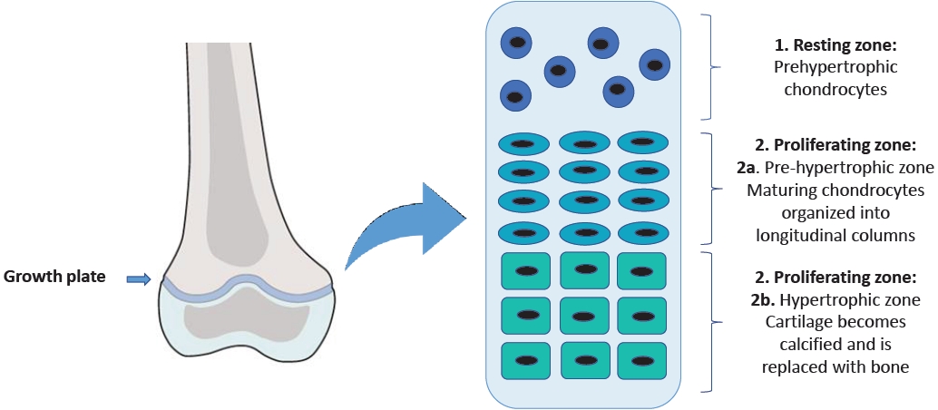
A wide range of factors may cause a child's reduced longitudinal growth, resulting in short stature in adulthood. Some of these factors influence chondrogenesis in the epiphyseal growth plate and affect the development of the skeleton, which is the main determinant of height [4]. The growth plate is the cartilaginous structure between the epiphysis and metaphysis where linear growth occurs [5].
In this region, there are 3 cell populations organized into 3 different zones with peculiar characteristics [6]. The resting zone is made up of round chondrocytes known as prehypertrophic chondrocytes. The proliferating zone is divided into the prehypertrophic zone, containing the maturing chondrocytes organized in longitudinal columns, and the hypertrophic zone, where the cartilage gradually goes through ossification (Fig. 1) [7].
While chondrocytes undergo this process of proliferation and differentiation, their endoplasmic reticulum (ER) synthesizes extracellular proteins, such as collagen, proteoglycans, hyaluronan, and many other specific proteins that represent the structural components of the extracellular matrix (ECM) [8,9]. ECM has a critical role in the structural support of chondrocytes and represents a medium in which signaling molecules and growth factors are able to spread through the avascular cartilage toward the target cells [8,10]. Therefore, it is obvious that mutations in genes that regulate the ECM may cause isolated short stature or severe skeletal dysplasia. Many genetic causes of growth disorders have already been established, but recent genomewide studies have highlighted new monogenic causes of growth disorders [4].
One mechanism that may lead to ECM alterations is the so-called "endoplasmic reticulum stress," which determines the accumulation of folded proteins [11] and can cause altered proliferation and differentiation of the chondrocytes of the growth plate with consequent skeletal diseases, such as chondrodysplasias [12].
Chondrodysplasias represent a heterogeneous group of rare genetic diseases caused by various etiologies involving more than 450 genes [13]; as a group, the incidence of chondrodysplasias is 1 per 5,000 births [14]. They are multisystem disorders whose main characteristic is represented by severe short stature, often associated with skeletal deformities, craniofacial anomalies, and premature joint degeneration. Additionally, these disorders may be associated with involvement of other organs, including neurological, auditory, visual, pulmonary, cardiac, renal, and psychological complications [14].
In this review, we aim to describe the main causes of short stature, focusing on the genes involved in ECM disorders (Table 1).

Collagens (COL2A1, COL10A1, COL9A2, COL11A1, COL27A)

Collagens represent one of the primary proteins in the ECM. Mutations in collagen genes are involved in many diseases like chondrodysplasias [15].
COL2A1 gene (12q13.1-q13.2) encodes type II collagen, a fibrillar protein of 1,487 amino acids. Type II collagen is the main component of hyaline cartilage ECM. In the growth plate, it is synthesized by the chondrocytes of the proliferation zone [16]. Type II collagen is composed of 3 identical α1-polypeptide chains made up of 1060 amino acids. Type II collagen molecules self-assemble into cross-linked fibrils which form the type II collagen fibers. These fibers interact with other macromolecules, forming the ECM of cartilage [17].
Mutations in the COL2A1 gene cause a wide variety of rare autosomal dominant diseases. More than 400 mutations have been described. These mutations are associated with an accumulation of immature procollagen in the cisternae of chondrocytes rough ER, causing cellular stress [18]. Furthermore, mutated type II collagen molecules would not be able to selfassemble into fibrils, which are necessary to allow for the correct columnar arrangement of the chondrocytes in the growth plate or to interact correctly with the other macromolecules of the matrix [19].
Mutations of this gene are responsible for spondyloepimetaphyseal dysplasia (SEMD) and spondyloepiphyseal dysplasia (SED) congenita, which are characterized by delayed ossification of the vertebrae and pubic bones with subsequent dwarfism, kyphoscoliosis in early childhood, and shortening of long bones. In spondyloperipheral dysplasia, patients show an important short stature (mean length 45 cm at term) associated with the absence of ossification in pubic bones, distal femoral epiphyses, and cervical and sacral vertebras with consequent lumbar lordosis.
COL10A1 (6q22.1) encodes collagen X, a homotrimer collagenous protein formed by 3 α-1 (X) chains. Type X collagen is expressed only in the hypertrophic chondrocytes layer of the cartilage growth plate.
Schmid's metaphyseal chondrodysplasia (SMCD) is inherited in autosomal dominant manner or with a novo mutation phenotype and seems to be related to an abnormal assembly of collagen trimer in the ER of hypertrophic chondrocytes, due to mutations in COL10A1 C-terminal noncollagen domain (NC1), which determines ER stress and reduced levels of functional type X collagen [20].
SMCD is typically diagnosed in early childhood and is characterized by progressive short stature starting from 2 years (adult height is usually more than 3.5 SD below the mean) associated with genu varum and waddling gait. Facial features and head size are normal, and there are no extraskeletal manifestations [21].
COL9A2 encodes type IX collagen, a heterotrimer fibrillar protein of the ECM. Each peptide chain consists of 3 collagenous domains separated by 4 noncollagenous domains [22]. Several mutations causing a loss of amino acids in the third collagenous domain are involved in the pathogenesis of multiple epiphyseal dysplasia (MED) [23].
MED is a skeletal dysplasia which, genetically, can be related to mutations in COMP, MATN3, COL9A1, COL9A2, and COL9A3, characterized by different clinical features. Clinical progress associated with COL9-MED is relatively mild, showing joint disease but only sometimes altered growth. Epiphyseal ossification delay and alterations in their shape are the main radiographic signs [24]. The MED phenotype usually manifests after the first 1–2 years of life. One of the first signs is joint pain, especially after exercise, affecting the hip and knee joints. On the contrary, growth failure is slowly progressive and can lead to mild to moderate short stature (approximately below the third percentile) at the age of 5–6 years [25].
COL11A1 and COL27A1 play a decisive role in the organization of the growth plate proliferative zone. Mutations in these proteins are associated with several forms of skeletal dysplasia [26-28]. Heterozygous mutations in the COL11A1 gene cause Stickler's syndrome [29] and Marshall's syndrome, which are phenotypically similar to each other, involving ophthalmic, articular, orofacial, and auditory defects. Radiographically, joint surface irregularities have been described [30,31]. Homozygous mutations of COL11A1 cause fibrochondrogenesis 1, a skeletal dysplasia presenting with short limbs, flat midface, and protrudent abdomen [32].
The COL27A1 gene encodes for collagen type XXVII, proalpha 1, and is located on human chromosome 9q32-33. The protein consists of 1860 amino acids and plays a pivotal role in the organization of ECM [33].
Recessive mutations in the COL27A1 gene are responsible for a rare genetic disorder called Steel syndrome [34,35], which is characterized by mesomelic short stature, bilateral genua valga, absence of capital femoral epiphyses, shallow bilateral acetabula, incomplete ossification of pubic bones, scoliosis, pectus excavatum, and facial dysmorphism [36].

Fibrillins

Fibrillins (FBN1) are structural proteins that, like collagen, form fibers and interact with many other connective tissue molecules [37]. In the human genome, 3 different genes (FBN1, FBN2, and BN3) encode fibrillins; FBN1 and FBN2 are mainly involved in growth regulation [38]. The human FBN1 gene is located on chromosome 15q15 21.1 and comprises 65 exons. FBN1 encodes fibrillin-1, a calcium-binding protein composed of 2871 amino acids that forms 10–12 nm microfibrils in the ECM [39].
Fibrillin microfibrils play a pivotal role in the structural integrity of many organs, such as the aortic wall and the suspensory ligament of the lens [37]. FBN1 mutation causes several genetic connective tissue diseases called fibrillinopathies [40], such as Marfan syndrome, an autosomal dominant disorder characterized by tall stature, skeletal and ocular abnormalities, cardiovascular involvement [41], and acromelic dysplasias characterized by short stature, short extremities, and joint stiffness. Acromelic dysplasias include Weill-Marchesani syndrome (WMS), geleophysic dysplasia, acromicric dysplasia (AD), and Myhre syndrome. The first condition in which FBN1 mutation has been observed was WMS [42], which is caused by an in-frame deletion of 24 nucleotides in exon 41 [43], while missense mutations in exons 41 and 42 cause geleophysic dysplasia and AD [44]. In few cases, an in-frame deletion of exons 9–11 may also be responsible for WMS [45].
Currently, 4 types of WMS have been described: type I, caused by a homozygous mutation in the ADAMTS10 gene; type II, caused by a heterozygous mutation in FBN1; and types III and IV, which are due to homozygous mutations in LTBP2 and ADAMTS17, respectively [43].
The primary clinical features of WMS are proportionate short stature, brachydactyly, joint stiffness, ocular abnormalities, and cardiac involvement [46]. Although some of these alterations such as ophthalmic abnormalities have been extensively described, there is still no detailed information in the literature regarding short stature and a possible correlation with growth hormone (GH) deficiency or whether these patients respond to GH therapy [46]. Short stature is reported in all affected individuals and appears in the first years of life. The average height of an adult male with WMS is approximately 142–169 cm and that of an adult female is 130–157 cm [47].
Geleophysic dysplasia is a severe form of acromelic dysplasia. Two types of geleophysic dysplasia have been described: type I, due to an ADAMTSL2 recessive mutation [48], and type II, caused by autosomal dominant mutations in FBN1 [49]. The term "geleophysic" derives from geleos, which means happy, and physis, which refers to nature. In fact, patients present with a "happy" face, with full cheeks, small palpebral fissures, short nose with anteverted nares, long smooth philtrum, and thin vermilion border of the upper lip [50]. Geleophysic dysplasia is characterized by short stature (<3 SD), brachydactyly, dysplastic femoral epiphysis, hepatomegaly, joint stiffness, progressive cardiac valvular thickening, and tracheal stenosis with bronchopulmonary insufficiency [51].
The main characteristics of AD are severe short stature, joint stiffness, shortened hands and feet, and mild facial dimorphism without cognitive impairment. Patients have normal height at birth but progressively decrease in centiles through childhood [44].

Aggrecan

Aggrecan (ACAN), the most important proteoglycan of growth plate cartilage, is encoded by the ACAN gene, which is located in chromosome 15q26.1 [52]. It is composed of an N-terminal domain, 2 globular domains (G1 and G2), 2 interglobular domains (CS and KS), a selectin-like domain (G3), and a C-terminal domain [53,54].
Aggrecan-type SEMD is caused by an autosomal recessive mutation in the G3 domain of aggrecan (p.Asp2267Asn) [55] and represents a severe skeletal dysplasia characterized by extreme short stature (final stature of 66–71 cm). Other clinical features include dysmorphic craniofacial abnormalities, brachydactyly, barrel chest, and mild lumbar lordosis [56]. Radiographic examination showed irregular epiphyses and enlarged metaphyses, particularly in the knees.
SED, Kimberley type, is a less severe skeletal dysplasia caused by an autosomal dominant mutation, resulting in the creation of a premature stop codon (p.Gly1330Trpfs * 221) [57]. It is characterized by proportionate short stature (<5th percentile; males 141–12 cm and females 136–149 cm) with stocky appearance and severe progressive osteoarthritis of the large ones [58]. Radiographic features include endplate irregularity and vertebral body sclerosis with mild and variable epiphyseal changes associated with delayed bone age [59].

Cartilage oligomeric matrix protein

Cartilage oligomeric matrix protein (COMP) is a homopentameric protein belonging to the thrombospondin gene family (TSP), whose gene is located on chromosome 19p12-13.1 [60]. The structure of TSP proteins has a common organization consisting of an N-terminal domain, 4 type 2 epidermal growth factor (EGF)-like repeats, 7 calcium-binding repeats of type 3, and a C-terminal globular domain [61,62]. Fare clic o toccare qui per immettere il testo.
The function of COMP would seem to be stimulation of chondrogenesis and proliferation of chondrocytes [63] and mediation of the interaction between proteins of the ECM in cartilage and other tissues [64,65]. More than 50 mutations have been identified, and most of these have been found in highly conserved repeat type 3 calcium-binding domains [66].
COMP mutations would be responsible for the accumulation of COMP and other ECM proteins within the ER of chondrocytes, causing a decrease in the proliferation of chondrocytes. COMP mutations are associated with 2 autosomal dominant skeletal dysplasias: pseudoachondroplasia (PSACH), a severe condition of dwarfism, and MED, a milder short stature disorder [67]. While all PSACH are related to COMP mutations, only 66% of MED are caused by COMP mutations [68].
PSACH is a condition of disproportionate dwarfism involving both the spine and limbs associated with joint abnormalities [69]. PSACH patients initially show normal growth, but begin to show the first signs of short stature by the end of the first year. The result is an adult height equivalent to that of an average 6-to 8-year-old child [70]. The face traits are distinctive and characteristic features. All joints are affected and are extremely lax. Joint pain is a major complication that begins in childhood and persists into adulthood [71].
Characteristics include radiographic findings of long bones shortening, enlarged and irregular metaphyses, small underossified femoral epiphyses, and platyspondyly [71].
MED is the milder skeletal dysplasia, presenting with epiphyseal abnormalities and joint pain which start in childhood [72]. MED patients are diagnosed around 5 years of age due to abnormal gait and/or joint stiffness and pain. Height is only slightly altered, with some patients achieving normal height. Early osteoarthritis, which primarily affects the hips, requires hip replacement in early adulthood [66].

Matrilin-3

Matrilin-3 (MATN3) is an ECM protein that is part of the matrilin family, whose gene is located on chromosome 2p24-p23. The matrilin family consists of 4 proteins formed by 1 or 2 Von Willebrand factor A (VWFA) domains, a variable number of EGF-like domains, and an α-helical spiral domain. MATN3 contains a VWFA domain, 4 EGF domains, and a C-terminal spiral domain [73,74]. The matrilins participate in the ECM assembly, and MATN3 mediates the interactions between collagen fibrils and the other ECM proteins [66,75]. Furthermore, MATN3 seems to play a role in chondrogenesis, premature maturation of chondrocytes, and ossification. In MATN3 knockout mice, growth plate chondrocytes transform early into a prehypertrophic and hypertrophic phenotype and form an expanded zone of hypertrophy [76]. MED can also be caused by MATN3 mutations [77,78] which cause retention of the protein within the ER, resulting in cellular stress [79,80].

A disintegrin and metalloprotease with thrombospondin motifs 17

A disintegrin and metalloprotease with thrombospondin motifs 17 (ADAMTS17) belongs to the ADAMTS family of ECM proteases, functionally related to fibrillins. It would seem that ADAMTS17 promotes the secretion and/or deposition of FBN1 and type I collagen in the ECM [81,82]. Furthermore, both in cell culture systems and in vivo, direct interactions among fibrillin-1, ADAMTS17, and LTBP2 have been demonstrated.
All ADAMTS proteases are characterized by a similar structure; they have a conserved N-terminal domain and a variable C-terminal auxiliary domain, which is involved in binding with other ECM proteins [83].
Mutations in ADAMTS17 are associated with WMS. Individuals with WMS type IV due to ADAMTS17 mutations present with predominantly musculoskeletal and ocular manifestations, having less severe cardiac involvement [82,84,85].
In genome-wide studies, the involvement of the ADAMTS17 locus is often correlated with human height [86]. Therefore, it is possible that familial short stature may be associated with suspected monoallelic ADAMTS17 deficiency. However, this hypothesis needs further investigation.

Latent transforming growth factor beta-binding proteins

LTBPs are important ECM proteins; 4 isoforms of latent transforming growth factor beta-binding proteins (LTBP2) interacting with fibrillin microfibrils have been identified in the human genome [87]. Electron microscopic examination revealed that LTBP2 could play a role in the formation of elastic fibers and in the assembly of extracellular microfibrils [39].
LTBP2 interacts with heparin and heparan sulfate proteoglycans through its N-terminal domain and, more weakly, through its central region [88]. These interactions would lead to further anchoring of the microfibrils to basal membranes.
Homozygous mutations in the LTBP2 gene cause WMS type III, primary congenital glaucoma [89], and appear to be involved in the pathogenesis of Marfan syndrome [90].

Fibronectin 1

Fibronectin 1 (FN1) (2q35) encodes for fibronectin [91]. Mutations in the FN1 gene impair cysteine residues that form disulfide bonds, on which depends the 3-dimensional structure of the protein, and their perturbation leads to instability and possible risk of degradation by metalloproteinases (MMP9 and MMP13) [92].
Mutations in the N-terminal domain, which is necessary for fibronectin interaction to form fibrils, lower the number of fibrils in the cell matrix [93]. Some pathogenic variants in the III-2 domain result in greatly reduced secretion of the protein and accumulation of abnormal fibronectin within the cells. Molecular genetic testing may identify some heterozygous mutations associated with spondylometaphyseal dysplasia, corner fracture type (SMDCF), a skeletal dysplasia characterized by short stature which may be present at birth or develop in early childhood. Individuals may present with short limbs and/or short trunk, and the final adult height is more than 2 SD below the mean. Some distinguishing features reported in individuals with FN1-SMDCF include nonspecific dysmorphic facial features and frequent fractures due to low bone mineral density. In addition, limited joint mobility and chronic pain are common.
Radiological characteristics include enlarged metaphyses with corner fracture-like lesions, coxa vara, shortened long bones, scoliosis, and vertebral anomalies (e.g., ovoid-shaped vertebral bodies, anterior wedging, narrow intervertebral spaces, vertebral fusion, vertebral hypoplasia) and joint stiffness with pain [94].

Conclusions

Short stature is one of the main reasons for a referral to a pediatric endocrinologist. In this review, we discussed the main causes of short stature, focusing on single genes defects involving the ECM of the growth plate. These mutations most often cause rare, complex, and multisystemic clinical pictures characterized by severe harmonic or disharmonious short stature. However, we have also observed that some variants of these mutations are frequently recognized in patients with idiopathic short stature who do not have syndromic clinical features. The new molecular genetics techniques will make it possible to recognize an increasing number of new genes and gene variants implicated in short stature, thereby allowing for the recognition of genetic short stature, which still remains misdiagnosed and identified as idiopathic forms.

Notes

Conflicts of interest

No potential conflict of interest relevant to this article was reported.

Funding

This study received no specific grant from any funding agency in the public, commercial, or not-for-profit sectors.

Author contribution

Writing - original draft: MAS, AQ, FC; Writing - review & editing: MASi, AQ, FC

Fig. 1.
The structure of the growth plate.
apem-2244120-060f1.jpg
Table 1.
Extracellular matrix genetic disorders involved in short stature
Gene Protein Disorders Clinical presentation
COL2A1 Collagen type II Spondyloepimetaphyseal dysplasia Dwarfism caused by delayed ossification of the vertebrae and pubic bones.
Spondyloepiphyseal dysplasia congenita Kyphoscoliosis develops in childhood
Spondyloperipheral dysplasia Short stature associated with important lumbar lordosis
COL10A1 Collagen type X Schmid metaphyseal chondrodysplasia Progressive short stature that develops by age 2 years, short limbs, genu varum, and waddling gait
COL9A2 Collagen type IX Multiple epiphyseal dysplasia Joint pain affecting the hip and knee joints, occurring initially, after physical exercise, progressive deviation from the normal growth curve, resulting in mild to moderate short stature
COL11A1 Collage type XI, α1-chain Stickler syndrome Marshall syndrome Ophthalmic (myopia, vitreoretinal degeneration, cataracts, and, often, retinal detachment), articular, orofacial, and auditory manifestations
Fibrochondrogenesis 1 Short limbs, flat midface and protrudent abdomen
COL27A1 Collagene type XXVII, α1-chain Steel Syndrome Short stature with mesomelic shortening and bilateral genua valga, absent capital femoral epiphyses, shallow bilateral acetabulae, incomplete ossification of pubic bones, scoliosis, pectus excavatum, and facial dysmorphism
FBN1 Fibrillin-1 Acromelic dysplasias: Proportionate short stature, brachydactyly, joint stiffness, ocular abnormalities and cardiac defects
-Weill-Marchesani syndrome type II “Happy” face, short stature, brachydactyly, small or dysplastic capital femoral epiphyses, hepatomegaly, limited joint mobility, and progressive cardiac valvular thickening
-Geleophysic dysplasia
-Acromicric dysplasia Severe shor t stature associated with joint stiffness and shortened hands and feet, normal intelligence, and mild facial dysmorphism
ACAN Aggrecan Spondyloepimetaphyseal dysplasia aggrecan type Severe skeletal dysplasia, extreme short stature with short necks, brachydactyly, barrel chest and mild lumbar lordosis, craniofacial abnormalities (macrocephaly, severe midface hypoplasia and low set ears)
Spondyloepiphyseal dysplasia Kimberley type Proportionate short stature (<5th percentile; males 141–162 cm and females 138–149 cm) with a stocky appearance and severe progressive osteoarthritis of the large weight bearing joints
COMP Cartilage oligomeric matrix protein Pseudoachondroplasia Disproportionate dwarfing, decelerating linear growth starting by the end of the first year, extremely lax joints, pain
Multiple epiphyseal dysplasia Epiphyseal abnormalities and joint pain starting in childhood
MTN-3 Matrilin-3 Multiple epiphyseal dysplasia Epiphyseal abnormalities and joint pain starting in childhood, osteoarthritis
ADAMTS17 Metalloproteases Weill-Marchesani syndrome type IV Less severe cardiac involvement, presents predominantly with the musculoskeletal and ocular features
LTBP2 Latent transforming growth factor beta-binding proteins Weill-Marchesani syndrome type III Short stature, brachydactyly, joint stiffness, ocular abnormalities and cardiac defects, thick skin
FN1 Fibronectin-1 Spondylometaphyseal dysplasia, corner fracture type Short stature, short limbs and/or short trunk. Radiographic features include enlargement and corner fracture-like lesions of the metaphyses, developmental coxa vara, shortened long bones, scoliosis, and vertebral anomalies. Limited joint mobility and chronic pain.

References

1. Bang P. Statement 3: a low serum IGF-I level in idiopathic short stature patients indicates partial GH insensitivity. Pediatr Endocrinol Rev 2008;5 Suppl 3:841–6.
pmid
2. Patel R, Dave C, Agarwal N, Mendpara H, Shukla R, Bajpai A. Predictive value of IAP 2015, IAP 2007 and WHO growth charts in identifying pathological short stature. Indian Pediatr 2021;58:149–51.
crossref pmid pdf
3. Chiarelli F, Primavera M, Mastromauro C. Evaluation and management of a child with short stature. Minerva Pediatr 2020;72:452–61.
crossref pmid
4. Yue S, Whalen P, Jee YH. Genetic regulation of linear growth. Ann Pediatr Endocrinol Metab 2019;24:2–14.
crossref pmid pmc pdf
5. Nilsson O, Marino R, De Luca F, Phillip M, Baron J. Endocrine regulation of the growth plate. Horm Res 2005;64:157–65.
crossref pmid pdf
6. Chen H, Tan XN, Hu S, Liu RQ, Peng LH, Li YM, et al. Molecular mechanisms of chondrocyte proliferation and differentiation. Front Cell Dev Biol 2021;9:664168.
crossref pmid pmc
7. Tsang KY, Tsang SW, Chan D, Cheah KS. The chondrocytic journey in endochondral bone growth and skeletal dysplasia. Birth Defects Res C Embryo Today 2014;102:52–73.
crossref pmid pdf
8. Faienza MF, Chiarito M, Brunetti G, D'Amato G. Growth plate gene involvement and isolated short stature. Endocrine 2021;71:28–34.
crossref pmid pdf
9. Schwartz NB, Domowicz M. Chondrodysplasias due to proteoglycan defects. Glycobiology 2002;12:57R–68R.
crossref pmid
10. Cortes M, Baria AT, Schwartz NB. Sulfation of chondroitin sulfate proteoglycans is necessary for proper Indian hedgehog signaling in the developing growth plate. Development 2009;136:1697–706.
crossref pmid pmc pdf
11. Rellmann Y, Dreier R. Different forms of ER stress in chondrocytes result in short stature disorders and degenerative cartilage diseases: new insights by cartilagespecific ERp57 knockout mice. Oxid Med Cell Longev 2018;2018:8421394.
crossref pmid pmc pdf
12. Patterson SE, Dealy CN. Mechanisms and models of endoplasmic reticulum stress in chondrodysplasia. Dev Dyn 2014;243:875–93.
crossref pmid
13. Krakow D. Skeletal dysplasias. Clin Perinatol 2015;42:301–19.
crossref pmid pmc
14. Krakow D, Rimoin DL. The skeletal dysplasias. Genet Med 2010;12:327–41.
crossref pmid
15. Myllyharju J, Kivirikko KI. Collagens and collagen-related diseases. Ann Med 2001;33:7–21.
crossref pmid
16. Lian C, Wang X, Qiu X, Wu Z, Gao B, Liu L, et al. Collagen type II suppresses articular chondrocyte hypertrophy and osteoarthritis progression by promoting integrin β1-SMAD1 interaction. Bone Res 2019;7:8.
crossref pmid pmc pdf
17. Antipova O, Orgel JP. In situ D-periodic molecular structure of type II collagen. J Biol Chem 2010;285:7087–96.
crossref pmid pmc
18. Prein C, Warmbold N, Farkas Z, Schieker M, Aszodi A, Clausen-Schaumann H. Structural and mechanical properties of the proliferative zone of the developing murine growth plate cartilage assessed by atomic force microscopy. Matrix Biol 2016;50:1–15.
crossref pmid
19. Chung HJ, Jensen DA, Gawron K, Steplewski A, Fertala A. R992C (p.R1192C) Substitution in collagen II alters the structure of mutant molecules and induces the unfolded protein response. J Mol Biol 2009;390:306–18.
crossref pmid pmc
20. Rajpar MH, McDermott B, Kung L, Eardley R, Knowles L, Heeran M, et al. Targeted induction of endoplasmic reticulum stress induces cartilage pathology. PLoS Genet 2009;5:e1000691.
crossref pmid pmc
21. Bateman JF, Freddi S, Nattrass G, Savarirayan R. Tissuespecific RNA surveillance? Nonsense-mediated mRNA decay causes collagen X haploinsufficiency in Schmid metaphyseal chondrodysplasia cartilage. Hum Mol Genet 2003;12:217–25.
crossref pmid
22. Pihlajamaa T, Vuoristo MM, Annunen S, Perälä M, Prockop DJ, Ala-Kokko L. Human COL9A1 and COL9A2 genes. Two genes of 90 and 15 kb code for similar polypeptides of the same collagen molecule. Matrix Biol 1998;17:237–41.
crossref pmid
23. Czarny-Ratajczak M, Lohiniva J, Rogala P, Kozlowski K, Perälä M, Carter L, et al. A mutation in COL9A1 causes multiple epiphyseal dysplasia: further evidence for locus heterogeneity. Am J Hum Genet 2001;69:969–80.
crossref pmid pmc
24. Unger S, Bonafé L, Superti-Furga A. Multiple epiphyseal dysplasia: clinical and radiographic features, differential diagnosis and molecular basis. Best Pract Res Clin Rheumatol 2008;22:19–32.
crossref pmid
25. Haga N, Nakamura K, Takikawa K, Manabe N, Ikegawa S, Kimizuka M. Stature and severity in multiple epiphyseal dysplasia. J Pediatr Orthop 1998;18:394–7.
crossref pmid
26. Plumb DA, Ferrara L, Torbica T, Knowles L, Mironov A Jr, Kadler KE, et al. Collagen XXVII organises the pericellular matrix in the growth plate. PLoS One 2011;6:e29422.
crossref pmid pmc
27. Hjorten R, Hansen U, Underwood RA, Telfer HE, Fernandes RJ, Krakow D, et al. Type XXVII collagen at the transition of cartilage to bone during skeletogenesis. Bone 2007;41:535–42.
crossref pmid pmc
28. Li Y, Lacerda DA, Warman ML, Beier DR, Yoshioka H, Ninomiya Y, et al. A fibrillar collagen gene, Col11a1, is essential for skeletal morphogenesis. Cell 1995;80:423–30.
crossref pmid
29. Van Camp G, Snoeckx RL, Hilgert N, van den Ende J, Fukuoka H, Wagatsuma M, et al. A new autosomal recessive form of Stickler syndrome is caused by a mutation in the COL9A1 gene. Am J Hum Genet 2006;79:449–57.
crossref pmid pmc
30. Annunen S, Körkkö J, Czarny M, Warman ML, Brunner HG, Kääriäinen H, et al. Splicing mutations of 54-bp exons in the COL11A1 gene cause Marshall syndrome, but other mutations cause overlapping Marshall/Stickler phenotypes. Am J Hum Genet 1999;65:974–83.
crossref pmid pmc
31. Griffith AJ, Sprunger LK, Sirko-Osadsa DA, Tiller GE, Meisler MH, Warman ML. Marshall syndrome associated with a splicing defect at the COL11A1 locus. Am J Hum Genet 1998;62:816–23.
crossref pmid pmc
32. Tompson SW, Bacino CA, Safina NP, Bober MB, Proud VK, Funari T, et al. Fibrochondrogenesis results from mutations in the COL11A1 type XI collagen gene. Am J Hum Genet 2010;87:708–12.
crossref pmid pmc
33. Pace JM, Corrado M, Missero C, Byers PH. Identification, characterization and expression analysis of a new fibrillar collagen gene, COL27A1. Matrix Biol 2003;22:3–14.
crossref pmid
34. Kotabagi S, Shah H, Shukla A, Girisha KM. Second family provides further evidence for causation of Steel syndrome by biallelic mutations in COL27A1. Clin Genet 2017;92:323–26.
crossref pmid pdf
35. Gariballa N, Ben-Mahmoud A, Komara M, Al-Shamsi AM, John A, Ali BR, et al. A novel aberrant splice site mutation in COL27A1 is responsible for Steel syndrome and extension of the phenotype to include hearing loss. Am J Med Genet A 2017;173:1257–63.
crossref pmid pdf
36. Evie Kritioti, Athina Theodosiou, Nayia Nicolaou, Angelos Alexandrou, Ioannis Papaevripidou, Elisavet Efstathiou, et al. First reported case of Steel syndrome in the European population: a novel homozygous mutation in COL27A1 and review of the literature. Eur J Med Genet 2020;63:103939.
crossref pmid
37. Sakai LY, Keene DR, Renard M, De Backer J. FBN1: the disease-causing gene for Marfan syndrome and other genetic disorders. Gene 2016;591:279–91.
crossref pmid pmc
38. Sakai LY, Keene DR. Fibrillin protein pleiotropy: acromelic dysplasias. Matrix Biol 2019;80:6–13.
crossref pmid
39. Robertson IB, Horiguchi M, Zilberberg L, Dabovic B, Hadjiolova K, Rifkin DB. Latent TGF-β-binding proteins. Matrix Biol 2015;47:44–53.
crossref pmid pmc
40. Cecchi A, Ogawa N, Martinez HR, Carlson A, Fan Y, Penny DJ, et al. Missense mutations in FBN1 exons 41 and 42 cause Weill-Marchesani syndrome with thoracic aortic disease and Marfan syndrome. Am J Med Genet A 2013;161A:2305–10.
crossref pmid pmc pdf
41. Cook JR, Ramirez F. Clinical, diagnostic, and therapeutic aspects of the Marfan syndrome. Adv Exp Med Biol 2014;802:77–94.
crossref pmid
42. Le Goff C, Cormier-Daire V. From tall to short: the role of TGFβ signaling in growth and its disorders. Am J Med Genet C Semin Med Genet 2012;160C:145–53.
crossref pmid
43. Faivre L, Gorlin RJ, Wirtz MK, Godfrey M, Dagoneau N, Samples JR, et al. In frame fibrillin-1 gene deletion in autosomal dominant Weill-Marchesani syndrome. J Med Genet 2003;40:34–6.
crossref pmid pmc
44. Le Goff C, Mahaut C, Wang LW, Allali S, Abhyankar A, Jensen S, et al. Mutations in the TGFβ binding-proteinlike domain 5 of FBN1 are responsible for acromicric and geleophysic dysplasias. Am J Hum Genet 2011;89:7–14.
crossref pmid pmc
45. Sengle G, Tsutsui K, Keene DR, Tufa SF, Carlson EJ, Charbonneau NL, et al. Microenvironmental regulation by fibrillin-1. PLoS Genet 2012;8:e1002425.
crossref pmid pmc
46. Al Motawa MNA, Al Shehri MSS, Al Buali MJ, Al Agnam AAM. Weill-Marchesani syndrome, a rare presentation of severe short stature with review of the literature. Am J Case Rep 2021;22:e930824.
pmid pmc
47. Marzin P, Cormier-Daire V, Tsilou E. Weill-Marchesani Syndrome. 2007 Nov 1 [updated 2020 Dec 10]. Adam MP, Mirzaa GM, Pagon RA, Wallace SE, Bean LJH, Gripp KW, et al., editors. GeneReviews® [Internet]. Seattle (WA): University of Washington, Seattle. 1993-2022.

48. Allali S, Le Goff C, Pressac-Diebold I, Pfennig G, Mahaut C, Dagoneau N, et al. Molecular screening of ADAMTSL2 gene in 33 patients reveals the genetic heterogeneity of geleophysic dysplasia. J Med Genet 2011;48:417–21.
crossref pmid pmc
49. Globa E, Zelinska N, Dauber A. The clinical cases of geleophysic dysplasia: one gene, different phenotypes. Case Rep Endocrinol 2018;2018:8212417.
crossref pmid pmc pdf
50. Kochhar A, Kirmani S, Cetta F, Younge B, Hyland JC, Michels V. Similarity of geleophysic dysplasia and weill-marchesani syndrome. Am J Med Genet A 2013;161A:3130–2.
crossref pmid pdf
51. Legare JM, Modaff P, Strom SP, Pauli RM, Bartlett HL. Geleophysic dysplasia: 48 year clinical update with emphasis on cardiac care. Am J Med Genet A 2018;176:2237–42.
crossref pmid pdf
52. Roughley PJ, Mort JS. The role of aggrecan in normal and osteoarthritic cartilage. J Exp Orthop 2014;1:8.
crossref pmid pmc pdf
53. Hauer NN, Sticht H, Boppudi S, Büttner C, Kraus C, Trautmann U, et al. Genetic screening confirms heterozygous mutations in ACAN as a major cause of idiopathic short stature. Sci Rep 2017;7:12225.
crossref pmid pmc pdf
54. Gkourogianni A, Andrew M, Tyzinski L, Crocker M, Douglas J, Dunbar N, et al. Clinical characterization of patients with autosomal dominant short stature due to aggrecan mutations. J Clin Endocrinol Metab 2017;102:460–9.
crossref pmid pdf
55. Gibson BG, Briggs MD. The aggrecanopathies; an evolving phenotypic spectrum of human genetic skeletal diseases. Orphanet J Rare Dis 2016;11:86.
crossref pmid pmc
56. Tompson SW, Merriman B, Funari VA, Fresquet M, Lachman RS, Rimoin DL, et al. A recessive skeletal dysplasia, SEMD aggrecan type, results from a missense mutation affecting the C-type lectin domain of aggrecan. Am J Hum Genet 2009;84:72–9.
crossref pmid pmc
57. Sentchordi-Montané L, Aza-Carmona M, Benito-Sanz S, Barreda-Bonis AC, Sánchez-Garre C, Prieto-Matos P, et al. Heterozygous aggrecan variants are associated with short stature and brachydactyly: description of 16 probands and a review of the literature. Clin Endocrinol (Oxf ) 2018;88:820–9.
crossref pmid pdf
58. Anderson IJ, Tsipouras P, Scher C, Ramesar RS, Martell RW, Beighton P. Spondyloepiphyseal dysplasia, mild autosomal dominant type is not due to primary defects of type II collagen. Am J Med Genet 1990;37:272–6.
crossref pmid
59. Gleghorn L, Ramesar R, Beighton P, Wallis G. A mutation in the variable repeat region of the aggrecan gene (AGC1) causes a form of spondyloepiphyseal dysplasia associated with severe, premature osteoarthritis. Am J Hum Genet 2005;77:484–90.
crossref pmid pmc
60. Posey KL, Hecht JT. The role of cartilage oligomeric matrix protein (COMP) in skeletal disease. Curr Drug Targets 2008;9:869–77.
crossref pmid
61. Bornstein P, Sage EH. Matricellular proteins: extracellular modulators of cell function. Curr Opin Cell Biol 2002;14:608–16.
crossref pmid
62. Adams JC, Lawler J. The thrombospondins. Cold Spring Harb Perspect Biol 2011;3:a009712.
crossref pmid pmc
63. Xu K, Zhang Y, Ilalov K, Carlson CS, Feng JQ, di Cesare PE, et al. Cartilage oligomeric matrix protein associates with granulin-epithelin precursor (GEP) and potentiates GEP-stimulated chondrocyte proliferation. J Biol Chem 2007;282:11347–55.
crossref pmid
64. Posey KL, Coustry F, Hecht JT. Cartilage oligomeric matrix protein: COMPopathies and beyond. Matrix Biology 2018;71-72:161–73.
crossref pmid pmc
65. Müller G, Michel A, Altenburg E. COMP (Cartilage Oligomeric Matrix Protein) is synthesized in ligament, tendon, meniscus, and articular cartilage. Connect Tissue Res 1998;39:233–44.
crossref pmid
66. Unger S, Hecht JT. Pseudoachondroplasia and multiple epiphyseal dysplasia: new etiologic developments. Am J Med Genet 2001;106:244–50.
crossref pmid
67. Briggs MD, Brock J, Ramsden SC, Bell PA. Genotype to phenotype correlations in cartilage oligomeric matrix protein associated chondrodysplasias. Eur J Hum Genet 2014;22:1278–82.
crossref pmid pmc pdf
68. Jackson GC, Mittaz-Crettol L, Taylor JA, Mortier GR, Spranger J, Zabel B, et al. Pseudoachondroplasia and multiple epiphyseal dysplasia: a 7-year comprehensive analysis of the known disease genes identify novel and recurrent mutations and provides an accurate assessment of their relative contribution. Hum Mutat 2012;33:144–57.
crossref pmid pmc pdf
69. Posey KL, Hayes E, Haynes R, Hecht JT. Role of TSP-5/COMP in pseudoachondroplasia. Int J Biochem Cell Biol 2004;36:1005–12.
crossref pmid
70. Mortier GR, Cohn DH, Cormier-Daire V, Hall C, Krakow D, Mundlos S, et al. Nosology and classification of genetic skeletal disorders: 2019 revision. Am J Med Genet A 2019;179:2393–419.
crossref pmid pdf
71. Gamble C, Nguyen J, Hashmi SS, Hecht JT. Pseudoachondroplasia and painful sequelae. Am J Med Genet A 2015;167A:2618–22.
crossref pmid
72. Briggs MD, Wright MJ, Mortier GR. Multiple epiphyseal dysplasia, autosomal dominant. 2003 Jan 8 [updated 2019 Apr 25]. Adam MP, Mirzaa GM, Pagon RA, Wallace SE, Bean LJH, Gripp KW, et al., editors. GeneReviews®[Internet]. Seattle (WA): University of Washington, Seattle. 1993-2022.

73. Klatt AR, Becker AKA, Neacsu CD, Paulsson M, Wagener R. The matrilins: modulators of extracellular matrix assembly. Int J Biochem Cell Biol 2011;43:320–30.
crossref pmid
74. Klatt AR, Nitsche DP, Kobbe B, Mörgelin M, Paulsson M, Wagener R. Molecular structure and tissue distribution of matrilin-3, a filament-forming extracellular matrix protein expressed during skeletal development. J Biol Chem 2000;275:3999–4006.
crossref pmid
75. Wagener R, Ehlen HWA, Ko YP, Kobbe B, Mann HH, Sengle G, et al. The matrilins - adaptor proteins in the extracellular matrix. FEBS Lett 2005;579:3323–9.
crossref pmid pdf
76. Pei M, Luo J, Chen Q. Enhancing and maintaining chondrogenesis of synovial fibroblasts by cartilage extracellular matrix protein matrilins. Osteoarthritis Cartilage 2008;16:1110–7.
crossref pmid pmc
77. Briggs MD, Chapman KL. Pseudoachondroplasia and multiple epiphyseal dysplasia: mutation review, molecular interactions, and genotype to phenotype correlations. Hum Mutat 2002;19:465–78.
crossref pmid
78. Posey KL, Yang Y, Veerisetty AC, Sharan SK, Hecht JT. Thrombospondins: from structure to therapeutics. Cell Mol Life Sci 2008;65:669–71.
crossref pmid pmc pdf
79. Nundlall S, Rajpar MH, Bell PA, Clowes C, Zeeff LAH, Gardner B, et al. An unfolded protein response is the initial cellular response to the expression of mutant matrilin-3 in a mouse model of multiple epiphyseal dysplasia. Cell Stress Chaperones 2010;15:835–49.
crossref pmid pmc
80. Leighton MP, Nundlall S, Starborg T, Meadows RS, Suleman F, Knowles L, et al. Decreased chondrocyte proliferation and dysregulated apoptosis in the cartilage growth plate are key features of a murine model of epiphyseal dysplasia caused by a matn3 mutation. Hum Mol Genet 2007;16:1728–41.
crossref pmid pmc
81. Wang LW, Kutz WE, Mead TJ, Beene LC, Singh S, Jenkins MW, et al. Adamts10 inactivation in mice leads to persistence of ocular microfibrils subsequent to reduced fibrillin-2 cleavage. Matrix Biol 2019;77:117–28.
crossref pmid pmc
82. Mularczyk EJ, Singh M, Godwin ARF, Galli F, Humphreys N, Adamson AD, et al. ADAMTS10-mediated tissue disruption in Weill–Marchesani syndrome. Hum Mol Genet 2018;27:3675–87.
crossref pmid pmc
83. Apte SS. A disintegrin-like and metalloprotease (reprolysintype) with thrombospondin type 1 motif (ADAMTS) superfamily: functions and mechanisms. J Biol Chem 2009;284:31493–7.
crossref pmid pmc
84. Faivre L, Dollfus H, Lyonnet S, Alembik Y, Mégarbané A, Samples J, et al. Clinical homogeneity and genetic heterogeneity in Weill-Marchesani syndrome. Am J Med Genet A 2003;123A:204–7.
crossref pmid
85. Khan AO, Aldahmesh MA, Al-Ghadeer H, Mohamed JY, Alkuraya FS. Familial spherophakia with short stature caused by a novel homozygous ADAMTS17 mutation. Ophthalmic Genet 2012;33:235–9.
crossref pmid
86. Gudbjartsson DF, Walters GB, Thorleifsson G, Stefansson H, Halldorsson BV, Zusmanovich P, et al. Many sequence variants affecting diversity of adult human height. Nat Genet 2008;40:609–15.
crossref pmid pdf
87. Hyytiäinen M, Taipale J, Heldin CH, Keski-Oja J. Recombinant latent transforming growth factor β-binding protein 2 assembles to fibroblast extracellular matrix and is susceptible to proteolytic processing and release. J Biol Chem 1998;273:20669–76.
crossref pmid
88. Parsi MK, Adams JRJ, Whitelock J, Gibson MA. LTBP-2 has multiple heparin/heparan sulfate binding sites. Matrix Biol 2010;29:393–401.
crossref pmid
89. Lewis CJ, Hedberg-Buenz A, DeLuca AP, Stone EM, Alward WLM, Fingert JH. Primary congenital and developmental glaucomas. Hum Mol Genet 2017;26(R1):R28–36.
crossref pmid pmc
90. Haji-Seyed-Javadi R, Jelodari-Mamaghani S, Paylakhi SH, Yazdani S, Nilforushan N, Fan JB, et al. LTBP2 mutations cause Weill-Marchesani and Weill-Marchesani-like syndrome and affect disruptions in the extracellular matrix. Hum Mutat 2012;33:1182–7.
crossref pmid
91. Cadoff EB, Sheffer R, Wientroub S, Ovadia D, Meiner V, Schwarzbauer JE. Mechanistic insights into the cellular effects of a novel FN1 variant associated with a spondylometaphyseal dysplasia. Clin Genet 2018;94:429–37.
crossref pmid pmc pdf
92. Costantini A, Valta H, Baratang NV, Yap P, Bertola DR, Yamamoto GL, et al. Novel fibronectin mutations and expansion of the phenotype in spondylometaphyseal dysplasia with “corner fractures. ” Bone 2019;121:163–71.
crossref
93. Lee CS, Fu H, Baratang N, Rousseau J, Kumra H, Sutton VR, et al. Mutations in fibronectin cause a subtype of spondylometaphyseal dysplasia with “corner fractures. ” Am J Hum Genet 2017;101:815–23.
crossref pmid pmc
94. England J, McFarquhar A, Campeau PM. Spondylometaphyseal dysplasia, corner fracture type. 2020. Adam MP, Mirzaa GM, Pagon RA, Wallace SE, Bean LJH, Gripp KW, et al., editors. GeneReviews® [Internet]. Seattle (WA): University of Washington, Seattle. 1993-2022.


ABOUT
ARTICLE CATEGORY

Browse all articles >

BROWSE ARTICLES
AUTHOR INFORMATION
Editorial Office
501-107, 30 Seocho-daero 74-gil, Seocho-gu, Seoul 06622, Republic of Korea
Tel: +82-2-3471-4268    Fax: +82-2-3471-4269    E-mail: editor@e-apem.org                

Copyright © 2024 by Korean Society of Pediatric Endocrinology.

Developed in M2PI

Close layer
prev next